Illustrated presentation
Contents
Lessons from the jellyfish’s green light
An unexpected catch for Shimomura
Tsien creates a palette with all the colours of the rainbow
A brilliant experiment by Chalfie
Martin Chalfie, Osamu Shimomura, Roger Y. Tsien
Further reading
Credits
The Royal Swedish Academy of Sciences has decided to award the Nobel Prize in Chemistry for 2008 jointly to Martin Chalfie, Osamu Shimomura and Roger Y. Tsien, “for the discovery and development of the green fluorescent protein, gfp”.
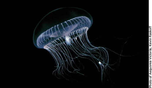
Lessons from the jellyfish’s green light
Off the west coast of North America, floats the jellyfish Aequorea victoria. In its light-emitting organs resides the green fluorescent protein, GFP, which glows intensely under ultraviolet light. GFP now revolutionizes the life sciences, and the scientists responsible for its development have been awarded this year’s Nobel Prize in Chemistry. The green light enables scientists to track, amongst other things, how cancer tumours form new blood vessels, how Alzheimer’s disease kills brain neurons and how HIV infected cells produce new viruses.
An unexpected catch for Shimomura
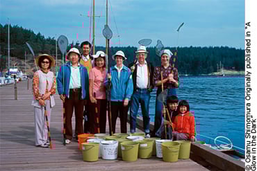
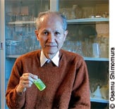
Throughout the summers of the 1960s, Osamu Shimomura (rearmost in the picture above), his family and various assistants, caught tens of thousands of jellyfish in the Pacific Ocean. From the edge, which emit green light when the jellyfish is agitated, Shimomura isolated a protein. Surprisingly, that protein did not shine in green. It was blue. Shimomura assumed that additional proteins were involved, and indeed found one more. It was not luminescent, but did glow bright green under the light of an ultraviolet lamp.
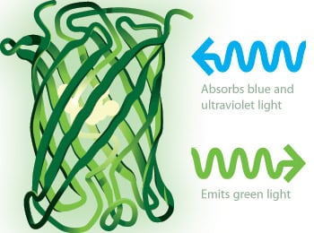 |
| A green guiding star for biosciences Today, scientists use GFP to understand the function of cells and proteins in living creatures. Proteins are the chemical tools of life – they control most of what happens within a living cell. Every human being functions thanks to the well-oiled machinery of thousands of proteins, like haemoglobin, antibodies and insulin. If something malfunctions, illness and disease often follows. Therefore it is fundamental for the biosciences to map out the role of various proteins. Using DNA-technology, scientists connect GFP to interesting, but otherwise invisible, proteins. GFP functions like a little lantern, which is activated by ultraviolet light. The green glow helps scientists track these proteins in the body. |
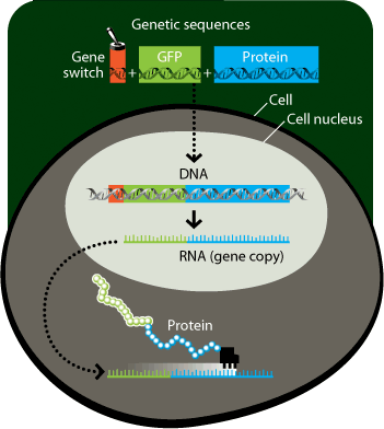
From Gene to Protein:
Tsien creates a palette with all the colours of the rainbow
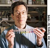
During the 1990s Roger Tsien explored and changed GFP. His playful research resulted in proteins that glowed cyan, blue and yellow. However, he did not manage to produce any red colours. Red light penetrates skin and other biological tissue more easily, and so is especially useful for research.
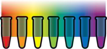 |
In 1999, Russian scientists isolated a red fluorescent protein, DsRED, from a coral. This protein was larger and more cumbersome than GFP. Tsien, however, managed to decrease the size of DsRED. From DsRED, Tsien also developed proteins with mouth-watering names like mPlum, mCherry, mStrawberry, mOrange and mCitrine.
A brilliant experiment by Chalfie
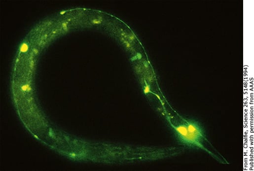
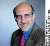
When Martin Chalfie first heard about GFP in 1988, he was delighted. He realised that GFP could possibly be used to colour cells and proteins. If that was the case, it would revolutionize the biosciences. The picture above shows Chalfie’s successful experiment. The touch receptor neurons of the millimetre-sized roundworm Caenorhabditis elegans fluoresces green. We can see the round bodies and the long slender projection of the nerve cells.
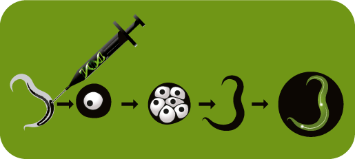
How cells become green Chalfie positioned the GFP-gene behind a promoter (a gene switch), which is active in the touch receptor neurons of the round worm. He injected the gene construct into the gonads of a mature worm. The worm is a hermaphrodite and fertilizes itself. The GFP gene is passed onto the eggs that the worm lays. The eggs divide, forming new individuals. The GFP-gene is then present in all cells of the new generation of roundworms, but only the touch receptor neurons will produce GFP. When they fill up with GFP, they start to glow green under ultraviolet light.
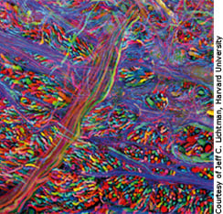
The brainbow
Scientists have used three fluorescent proteins in cyan, yellow and red – colours similar to those used by a computer printer – to colour the brain of a mouse. Different neurons randomly produce different amounts of the proteins. We can distinguish single neurons interlaced within the dense network.
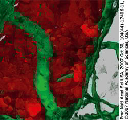
Tumour surrounded by nourishing blood vessels
Scientists have coloured a breast cancer tumour with DsRED and the surrounding blood vessels with GFP. In this experiment, scientists discovered two proteins which help breast cancer cells spread. If scientists can neutralize these proteins, they might also be able to stop the cells from breaking away from the tumour area.
 |
 |
 |
Martin Chalfie
US citizen. Born 1947 in Chicago, IL, USA. Professor of Biological Sciences at Columbia University, New York, NY, USA.
Osamu Shimomura
Japanese citizen. Born 1928 in Kyoto, Japan. Professor Emeritus at Marine Biological Laboratory (MBL), Woods Hole, MA, USA, and Boston University Medical School, MA, USA.
Roger Y. Tsien
US citizen. Born 1952 in New York, NY, USA. Professor and Investigator at Howard Hughes Medical Institute, University of California, San Diego, La Jolla, CA, USA.
Further reading
More information about the Nobel Prize in Chemistry for 2008 can be found on: www.kva.se, http://nobelprize.org, www.nobelmuseet.se
Pieribone, V. and Gruber, D.F. (2005) Aglow in the Dark, The Belknap Press, Harvard University Press, 2005
Zimmer, M. (2005) Glowing Genes, Prometheus Books
Shimomura, O. (2005) The discovery of aequorin and green fluorescent protein, Journal of Microscopy, 217 3-15
Shaner, N.C. et al (2008) Improving the photostability of bright monomeric orange and red fluorescent proteins, Nature Methods, 5 545-551
Movie showing a cell producing GFP-tagged HIV particles: www.nature.com/nature/journal/v454/n7201/suppinfo/nature06998.html
Website describing the GFP-revolution: http://www.conncoll.edu/ccacad/zimmer/GFP-ww/GFP-1.htm
Credits and references for the 2008 Nobel Poster for Chemistry
Editors: Gunnar von Heijne, Chairman of the Nobel Committee for Chemistry, the Royal Swedish Academy of Sciences, Ann Fernholm, Katalys Media, Annika Moberg and Andrea Westerdahl, the Royal Swedish Academy of Sciences.
Layout and illustrations by Typoform
Printed by AlfaPrint AB, Stockholm, 2008
Copyright © 2008 The Royal Swedish Academy of Sciences
Box 50005, SE-104 05 Stockholm, Sweden
Phone:+46 8 673 95 00, fax: +46 8 15 56 70
e-mail: [email protected], www.kva.se
Posters may be ordered free of charge by phone, fax or e-mail.
Web adapted version: Nobelprize.org
Nobel Poster from the Nobel Committee for Chemistry, web adapted by Nobel Web
Nobel Prizes and laureates
Six prizes were awarded for achievements that have conferred the greatest benefit to humankind. The 14 laureates' work and discoveries range from quantum tunnelling to promoting democratic rights.
See them all presented here.

