Press release
English
Swedish
French
German

NOBELFÖRSAMLINGEN KAROLINSKA INSTITUTET
THE NOBEL ASSEMBLY AT THE KAROLINSKA INSTITUTE
9 October 2000
The Nobel Assembly at Karolinska Institutet has today decided to award The Nobel Prize in Physiology or Medicine for 2000
Arvid Carlsson, Paul Greengard and Eric Kandel
for their discoveries concerning “signal transduction in the nervous system”
Summary
In the human brain there are more than hundred billion nerve cells. They are connected to each other through an infinitely complex network of nerve processes. The message from one nerve cell to another is transmitted through different chemical transmitters. The signal transduction takes place in special points of contact, called synapses. A nerve cell can have thousands of such contacts with other nerve cells.
The three Nobel Laureates in Physiology or Medicine have made pioneering discoveries concerning one type of signal transduction between nerve cells, referred to as slow synaptic transmission. These discoveries have been crucial for an understanding of the normal function of the brain and how disturbances in this signal transduction can give rise to neurological and psychiatric diseases. These findings have resulted in the development of new drugs.
Arvid Carlsson, Department of Pharmacology, Göteborg University is rewarded for his discovery that dopamine is a transmitter in the brain and that it has great importance for our ability to control movements. His research has led to the realization that Parkinson’s disease is caused by a lack of dopamine in certain parts of the brain and that an efficient remedy (L-dopa) for this disease could be developed. Arvid Carlsson has made a number of subsequent discoveries, which have further clarified the role of dopamine in the brain. He has thus demonstrated the mode of action of drugs used for the treatment of schizophrenia.
Paul Greengard, Laboratory of Molecular and Cellular Science, Rockefeller University, New York, is rewarded for his discovery of how dopamine and a number of other transmitters exert their action in the nervous system. The transmitter first acts on a receptor on the cell surface. This will trigger a cascade of reactions that will affect certain “key proteins” that in turn regulate a variety of functions in the nerve cell. The proteins become modified as phosphate groups are added (phosphorylation) or removed (dephosphorylation), which causes a change in the shape and function of the protein. Through this mechanism the transmitters can carry their message from one nerve cell to another.
Eric Kandel, Center for Neurobiology and Behavior, Columbia University, New York, is rewarded for his discoveries of how the efficiency of synapses can be modified, and which molecular mechanisms that take part. With the nervous system of a sea slug as experimental model he has demonstrated how changes of synaptic function are central for learning and memory. Protein phosphorylation in synapses plays an important role for the generation of a form of short term memory. For the development of a long term memory a change in protein synthesis is also required, which can lead to alterations in shape and function of the synapse.
Arvid Carlsson
Dopamine – an important transmitter
Arvid Carlsson performed a series of pioneering studies during the late 1950’s, which showed that dopamine is an important transmitter in the brain. It was previously believed that dopamine was only a precursor of another transmitter, noradrenaline. Arvid Carlsson developed an assay that made it possible to measure tissue levels of dopamine with high sensitivity. He found that dopamine was concentrated in other areas of the brain than noradrenaline, which led him to the conclusion that dopamine is a transmitter in itself. Dopamine existed in particularly high concentrations in those parts of the brain, called the basal ganglia, which are of particular importance for the control of motor behavior.
In a series of experiments Arvid Carlsson used a naturally occurring substance, reserpine, which depletes the storage of several synaptic transmitters. When it was given to experimental animals they lost their ability to perform spontaneous movements. He then treated the animals with L-dopa, a precursor of dopamine, which is transformed to dopamine in the brain. The symptoms disappeared and the animals resumed their normal motor behavior. In contrast, animals that received a precursor of the transmitter serotonin did not improve the motor behavior. Arvid Carlsson also showed that the treatment with L-dopa normalized the levels of dopamine in the brain.
Drugs against Parkinson’s disease
Arvid Carlsson realized that the symptoms caused by reserpine were similar to the syndrome of Parkinson’s disease. This led, in turn, to the finding that Parkinson patients have abnormally low concentrations of dopamine in the basal ganglia. As a consequence L-dopa was developed as a drug against Parkinson’s disease and today still is the most important treatment for the disease. During Parkinson’s disease dopamine producing nerve cells in the basal ganglia degenerate, which causes tremor, rigidity and akinesia. L-dopa, which is converted to dopamine in the brain, compensates for the lack of dopamine and normalizes motor behavior.
Antipsychotic and antidepressive drugs
Apart from the successful treatment of Parkinson’s disease Arvid Carlsson’s research has increased our understanding of the mechanism of several other drugs. He showed that antipsychotic drugs, mostly used against schizophrenia, affect synaptic transmission by blocking dopamine receptors. The discoveries of Arvid Carlsson have had great importance for the treatment of depression, which is one of our most common diseases. He has contributed strongly to the development of selective serotonin uptake blockers, a new generation of antidepressive drugs.
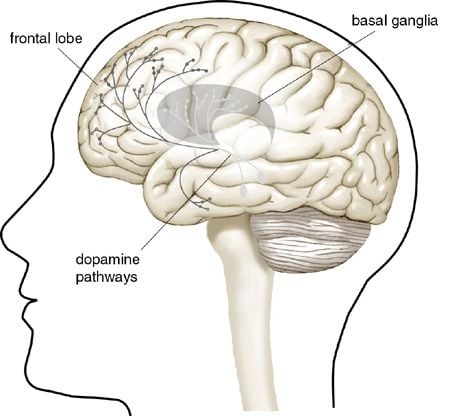 |
| Figure 1. Dopamine nerve pathways in the brain. Arvid Carlsson showed that there were particularly high levels of the chemical transmitter dopamine in the so called basal ganglia of the brain, which are of major importance for instance for the control of our muscle movements. In Parkinson’s disease those dopamine producing nerve cells whose nerve fibers project to the basal ganglia die. This causes symptoms such as tremor, muscle rigidity and a decreased ability to move about. |
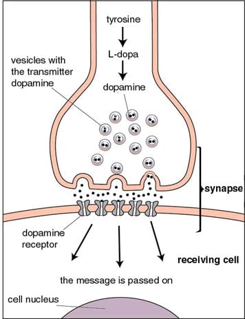
Figure 2.
A message from one nerve cell to another is transmitted with the help of different chemical transmitters. This occurs at specific points of contact, synapses, between the nerve cells. The chemical transmitter dopamine is formed from the precursors tyrosine and L-dopa and is stored in vesicles in the nerve endings. When a nerve impulse causes the vesicles to empty, dopamine receptors in the membrane of the receiving cell are influenced such that the message is carried further into the cell. In the treatment of Parkinson’s disease, the drug L-dopa is given, and is converted to dopamine in the brain. This compensates for the patient’s lack of dopamine.
Paul Greengard
Slow synaptic transmission
Towards the end of the 1960’s it was known that dopamine, noradrenaline and serotonin were transmitters in the central nervous system but knowledge about their mechanism of action was lacking. Paul Greengard receives the Nobel Prize for his discoveries of how they exert their effects at the synapse.
Transmitters such as dopamine, noradrenaline, serotonin and certain neuropeptides transmit their signals by what is referred to as slow synaptic transmission. The resulting change in the function of the nerve cell may last from seconds to hours. This type of signal transmission is responsible for a number of basal functions in the nervous system and is of importance for e.g. alertness and mood. Slow synaptic transmission can also control fast synaptic transmission, which in turn enables e.g. speech, movements and sensory perception.
Phosphorylation of proteins changes the function of nerve cells
Paul Greengard showed that slow synaptic transmission involves a chemical reaction called protein phosphorylation. It means that phosphate groups are coupled to a protein in such a way that the form and function of the protein is altered. Paul Greengard showed that when dopamine stimulates a receptor in the cell membrane this causes an elevation of a second messenger, cyclic AMP, in the cell. It activates a Protein Kinase A, which is able to add phosphate molecules to other proteins in the nerve cell.
The protein phosphorylation affects a series of proteins with different functions in the nerve cell. One important group of such proteins form ion channels in the membrane of the cell. They control the excitability of the nerve cell and make it possible for the nerve cell to send electrical impulses along its axons and terminals. Each nerve cell has different ion channels, which determine the reaction of the cell. When a particular type of ion channel is phosphorylated the function of the nerve cell may be altered by, for example, a change in its excitability.
DARPP-32 – a central regulatory protein
Paul Greengard has subsequently shown that even more complicated reactions occur in particular nerve cells. The effects of the transmitters are elicited by a cascade of phosphorylations and dephosphorylations (that is, phosphate molecules are added or removed from the proteins). Dopamine and several other transmitters can influence a regulatory protein, DARPP-32, which indirectly changes the function of a large number of other proteins. The DARPP-32 protein is like a conductor directing a series of other molecules. When DARPP-32 is activated it affects several ion channels altering the function of particular fast synapses.
Paul Greengard’s discoveries concerning protein phosphorylation have increased our understanding of the mechanism of action of several drugs, which specifically affects the phosphorylation of proteins in different nerve cells.
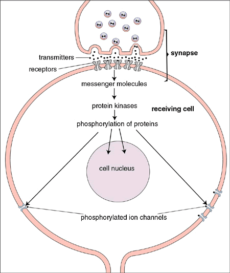 |
| Figure 3. Paul Greengard has shown how dopamine and several other chemical transmitters exert their effects in the nerve cell. When receptors in the cell membrane are influenced by a chemical transmitter, the levels of for example the messenger molecule cAMP are elevated. This activates so called protein kinases, which cause certain “key proteins” to become phosphorylated, that is phosphate molecules are added. These protein phosphorylations lead to changes of a number of proteins with different functions in the cell. When for instance proteins in ion channels in the cell membrane are influenced, the excitability of a nerve cell and its ability to send impulses along its branches changes. |
Eric Kandel
Sea slug, a model system for learning
A phosphorylation of proteins has great importance also for the discoveries for which Eric Kandel is rewarded, that is for revealing molecular mechanisms, important for the formation of memories. Eric Kandel started to study learning and memory in mammals, but realized that the conditions were too complex to provide an understanding of basic memory processes. He therefore decided to investigate a simpler experimental model, the nervous system of a sea slug, Aplysia. It has comparatively few nerve cells (around 20.000), many of which are rather large. It has a simple protective reflex that protects the gills, which can be utilized to study basic learning mechanisms.
Eric Kandel found that certain types of stimuli resulted in an amplification of the protective reflex of the sea slug. This strengthening of the reflex could remain for days and weeks and was thus a form of learning. He could then show that learning was due to an amplification of the synapse that connects the sensory nerve cells to the nerve cells that activate the muscle groups that give rise to the protective reflex.
Short and long term memory
Eric Kandel showed initially that weaker stimuli give rise to a form of short term memory, which lasts from minutes to hours. The mechanism for this “short term memory” is that particular ion channels are affected in such a manner that more calcium ions will enter the nerve terminal. This leads to an increased amount of transmitter release at the synapse, and thereby to an amplification of the reflex. This change is due to a phosphorylation of certain ion channel proteins, that is utilizing the molecular mechanism described by Paul Greengard.
A more powerful and long lasting stimulus will result in a form of long term memory that can remain for weeks. The stronger stimulus will give rise to increased levels of the messenger molecule cAMP and thereby protein kinase A. These signals will reach the cell nucleus and cause a change in a number of proteins in the synapse. The formation of certain proteins will increase, while others will decrease. The final result is that the shape of the synapse can increase and thereby create a long lasting increase of synaptic function. In contrast to short term memory, long term memory requires that new proteins are formed. If this synthesis of new proteins is prevented, the long term memory will be blocked but not the short term memory.
Synaptic plasticity, a precondition for memory
Eric Kandel thus demonstrated that short term memory, as well as long term memory in the sea slug is located at the synapse. During the 1990’s he has also carried out studies in mice. He has been able to show that the same type of long term changes of synaptic function that can be seen during learning in the sea slug also applies to mammals.
The fundamental mechanisms that Eric Kandel has revealed are also applicable to humans. Our memory can be said to be “located in the synapses” and changes in synaptic function are central, when different types of memories are formed. Even if the road towards an understanding of complex memory functions still is long, the results of Eric Kandel has provided a critical building stone. It is now possible to continue and for instance study how complex memory images are stored in our nervous system, and how it is possible to recreate the memory of earlier events. Since we now understand important aspects of the cellular and molecular mechanisms which make us remember, the possibilities to develop new types of medication to improve memory function in patients with different types of dementia may be increased.
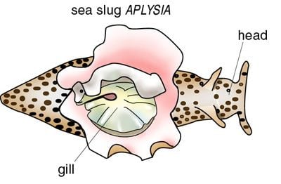
Figure 4.
A sea slug, Aplysia, has a simple nervous system and a gill withdrawal reflex that Eric Kandel has utilized to study learning and memory.
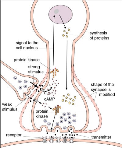 |
| Figure 5. A schematic description of how molecular changes in a synapse may produce “short term memory” and “long term memory” in the sea slug, Aplysia. The figure shows a synapse that is affecting another synapse. Short term memory can be produced when a weak stimulus (thin arrows in the left lower part of the figure) is causing a protein phosphorylation of ion channels, which leads to a release of an increased amount of transmitter. For a long term memory to be created, a stronger and more long-lasting stimulus is required (bold arrows in the figure). This causes an increased level of the messenger molecule cAMP, which causesa further activation of protein kinases. They will phosphorylate different proteins and affect the cell nucleus, which in turn will issue orders regarding the synthesis of new proteins. This may lead to changes in the form and function of the synapse. The efficacy of the synapse can then be increased and more transmitter released. |
Nobel Prizes and laureates
Six prizes were awarded for achievements that have conferred the greatest benefit to humankind. The 14 laureates' work and discoveries range from quantum tunnelling to promoting democratic rights.
See them all presented here.
