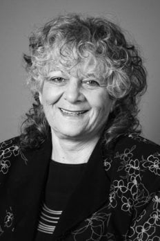
Ada E. Yonath
Biographical

I was born in Jerusalem in 1939 to a poor family that shared a rented fourroom apartment with two additional families and their children. My memories from my childhood are centered on my father’s medical conditions alongside my constant desire to understand the principles of the nature around me. The hard conditions didn’t dampen my enormous curiosity. Already at five, I was actively investigating the world. In one of my experiments I tried to measure the height of our tiny balcony using the furniture from inside the apartment. I put a table on another table, and then a chair and a stool on top, but did not reach the ceiling. Hence, I climbed up on my construct, fell down to the back yard on the ground floor and broke my arm … Incidentally, the results of this experiment are still unknown, since the current tenants in the apartment have remodeled the ceiling.
My parents were raised in religious families, being educated mainly in Judaism (my father) and women household skills (my mother). All of the schools in my immediate neighborhood were based on the same principles as those of my parents. However, despite the poverty of my parents and the lack of formal education, they went out of their way so that I could obtain a proper education in a very prestigious secular grammar school, called “Beit Hakerem”.
My father was frequently hospitalized and operated on, and when I was 11 years old he died. My mother barely coped, and I started to help her at that age. I had all types of jobs, cleaning, babysitting and providing private tuition to younger children. But both of us could not earn enough to support our little family, and consequently a year later my mother decided to move to another city, Tel Aviv, in order to be closer to her sisters. There I completed my high school education, and my mother, despite her tough life, supported my desire to keep on learning.
Indeed, after I spent my compulsory army service in the “top secret office” of the Medical Forces, where I was fortunate to be exposed to clinical and medical issues, I enrolled to the Hebrew University of Jerusalem. There I completed my undergraduate and M.Sc. studies in chemistry, biochemistry and biophysics. My doctoral work was carried out at the Weizmann Institute. I tried to reveal the high resolution structure of collagen. I continued to work on fibrous proteins (muscle) in my first postdoctoral year at the Mellon Institute in Pittsburg, Pennsylvania and then moved to the Massachusetts Institute of Technology (MIT) to study the structure of a globur protein staphylococcus nuclease. After completing my postdoctoral research, at the end of 1970, I returned to the Weizmann Institute. There, I initiated and established the first biological crystallography laboratory in Israel, which for almost a decade was the only laboratory for such studies.
At the end of the 1970s, I was a young researcher at the Weizmann Institute with an ambitious plan to shed light on one of the major outstanding questions concerning living cells: the process of protein biosynthesis. For this aim I wanted to determine the three-dimensional structure of the ribosome – the cells’ factory for translating the instructions written in the genetic code into proteins – and thus reveal the mechanics guiding the process. This was the beginning of a long quest that took over two decades, in which I was met with reactions of disbelief and even ridicule in the international scientific community. I can compare this journey to climbing Mt. Everest only to discover that a higher Everest stood in front of us.
I began these studies in collaboration with Prof. H.G. Wittmann of the Max Planck Institute for Molecular Genetics in Berlin, who supported these studies academically and financially. In parallel I maintained my laboratory at the Weizmann Institute, initially with a very modest budget and a grant given by the USA National Institute of Health (NIH). Over the years, a center for macromolecular assemblies was established by Mrs. Helen Kimmel at the Weizmann Institute, and consequently I came to lead a large team of researchers from all corners of the globe. Though my research began as an attempt to understand one of the fundamental components of life, it has led to a detailed understanding of the actions of some of the most widely prescribed antibiotics. My findings may not only aid in the development of more efficient antibacterial drugs, but could give scientists new weapons in the fight against antibiotic resistant bacteria – a problem that has been called one of the most pressing medical challenges of the 21st century.
Because the ribosome is so central to life, scientists around the world had been trying for many years to figure out how it works, but without an understanding of its spatial structure there was little hope of forming a comprehensive picture. To reveal the three-dimensional structure at the molecular level, crystals are required, but when dealing with ribosomes, there are added challenges. The ribosome is a complex of proteins and RNA chains; its structure is extraordinarily intricate; it is unusually flexible, unstable and lacks internal symmetry, all making crystallization an extremely formidable task.
At the start of the 1980s, working at both the Weizmann Institute in Israel and the Max Planck Institute in Germany, we created the first ribosome micro crystals. The procedure, which I developed especially for this aim, included a method for the preparation of the crystallizable ribosome that had been developed at the Weizmann Institute by Prof. Ada Zamir, Ruth Miskin and David Ellison. My inspiration came from an article on hibernating bears that pack their ribosomes in an orderly way in their cells just before hibernation, and these stay intact and potentially functional for months. Assuming that this is a natural strategy to maintain ribosomal activity for long time, I searched for ribosomes from organisms that live under harsh conditions, first of semi thermophiles, given by Dr. V. Erdmann and later I developed a unique experimental system based on ribosomes taken from the hardy bacteria living the extreme environments of the Dead Sea, thermal springs and atomic piles. In this way we managed to produce the initial micro crystals of ribosome in a fairly short time. However, even after obtaining preliminary diffraction indications, when I described my plans to determine the ribosome structure many distinguished scientists responded with sarcasm and disbelief. Consequently I became the World’s dreamer, the village fool, the so-called scientist, and the person driven by fantasies.
In the mid-1980s we visualized a tunnel spanning the large ribosomal subunit and assumed, based on previous biochemical works (Malkin & Rich, 1967, Blobel & Sabatini, 1970) that this is the path through which the nascent protein progresses as it is being formed – until it emerges out of the ribosome. In the course of my research, I developed a number of new techniques that are today widely used in structural biology labs around the world. One of these is cryo-bio-crystallography, which involves exposing the crystal to extremely low temperatures, –185°C, to minimize the crystalline structure’s disintegration under the X-ray bombardment. The day we conducted this experiment was special and unique. One of the rare “Eureka!” events. In retrospect, it was second only to the great pleasure I had when seeing our first high resolution structure a dozen years later. In fact the “Eureka type” of an experiment was not common, although we frequently had a great pleasure of overcoming complicated challenges.
In the mid-1990s, once we proved the feasibility of ribosome crystallography, several groups from leading universities or research institutions initiated parallel efforts. As they could repeat our procedures, I was no more alone in this field. At the end of the 1990s, we as well as those who used our experimental systems succeeded in breaking the resolution barrier, thanks to improvements in the crystals, in the facilities for detecting the X-ray diffraction and in ways to determine the diffraction phases. The first electron density map of the ribosome’s small subunit was a real breakthrough, and for me, a tremendous excitement. Then, in 2000 and 2001, we published the first complete three-dimensional structures of both subunits of the bacterial ribosome.
These discoveries are clearly a high point in 20 years of research, but my quest to understand the ribosome is still far from complete. Armed with new insight into ribosomal structure, I can afford moving on to revealing what else these structures can tell us about the ribosome actions, and how antibiotic drugs block those actions in bacterial ribosomes. Because ribosomes are so essential to life, many antibiotic drugs work by targeting their actions. The advances we made in our long quest to solve the structure and function of the ribosome may also pave the way toward improving existing antibiotic drugs or designing novel ones. We therefore crystallized bacterial ribosomes that can serve as pathogen models, complexed with each of over two dozens antibiotic compound. We found that the drugs bind in specific “pockets” in the structure, located at or close to functional centers, thus can block them and prevent the ribosomes from manufacturing proteins. Since these findings were published in Nature, in 2001, we have revealed the means of action of almost all of the antibiotics that target the ribosome, and our research in this area is ongoing.
For all scientists, the true scientific discovery is the top. In my case I can recall saying things like: ‘why work on ribosomes, they are dead … we know all what can be known about them’, or: ‘this is a dead end road’, or: ‘you will be dead before you get there’. Indeed, to my satisfaction, these predictions were proven wrong, the ribosomes are alive and kicking (so am I) and their high resolution structures stimulated many advanced studies.
And in the future? We plan on looking to the distant past. Ribosomes are found in every living being – from yeast and bacteria to mammals – and the structures of their active sites have been extraordinarily well-preserved throughout evolution. We have identified a region within the contemporary ribosome that seems to be the vestige of the primordial apparatus for producing peptide bonds and essentially giving rise to life. How did these first ribosomes come into being? How did they begin to produce proteins? How did they evolve into the sophisticated protein factories we see today in living cells? We plan on answering these and related questions in our future work.
Awarding the Nobel Prize exposed the ribosome to the public. It stimulated true scientific interest and turned on the imagination of many youngsters. As I have curly hair, there is a new saying in Israel: “Curly hair means a head full of ribosomes”. Furthermore, our studies added to the buzz around the lovely North Pole Bears, which inspired my own research and are now endangered by the changing climate.
These studies could not be performed without the help and/or active participation of many individuals. Thanks are due to the Weizmann Institute, particularly its presidents Michael Sela and Haim Harari, for keeping up with me for over two decades and for allowing me to work; to the Max Planck Society, especially the late Prof H.G. Wittmann for co-initiating this project, for producing the ribosome and their crystals and for financing my dream; to Ms. Helen Kimmel for establishing and maintaining the Kimmelman Center, thus paving the road for us from the early stages of our studies; to my colleagues in Hamburg (e.g. Frank Schluenzen, Heike Bartels, Joerg Harms and Ante Tocilj) and in Israel (especially Anat Bashan, Ilana Agmon, Tamar Awerbach, Ziva Berkovitch-Yellin, Raz Zarivach and Shulamit Weinstein), as well as my collaborators in Berlin (especially Francois Franceschi) for their devotion and enthusiasm in good and bad periods.
Above all, to my family who supported me with no questions or complaints despite my frequent disappearances and although at times my mind was not solely with them. These include my parents, who were brought up far away from science, especially my mother, who experienced enormous difficulties in raising and educating me after my dad’s death when I was still a child; my young sister Nurit, and my daughter Hagith, who had to tolerated me in my presence as well as in my absence; and to my granddaughter Noa, who at the age of five invited me to her kindergarten to talk about the ribosome!
This autobiography/biography was written at the time of the award and later published in the book series Les Prix Nobel/ Nobel Lectures/The Nobel Prizes. The information is sometimes updated with an addendum submitted by the Laureate.
Nobel Prizes and laureates
Six prizes were awarded for achievements that have conferred the greatest benefit to humankind. The 12 laureates' work and discoveries range from proteins' structures and machine learning to fighting for a world free of nuclear weapons.
See them all presented here.
