Brian Kobilka
Biographical
Growing Up In Little Falls, Minnesota
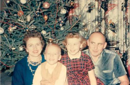
I was the second of two children born to Betty (Elisabeth) and Franklyn Kobilka on May 30, 1955 in Little Falls, Minnesota, a town of approximately 7,000 inhabitants along the Mississippi River. While small by most standards, Little Falls was the largest town in Morrison County and was known as the boyhood home of the pioneering aviator Charles Lindbergh. My sister Pam was three years older (Figure 1). My father owned and operated a bakery in Little Falls.
My father’s bakery was relatively large for a small town, with 6–8 full time employees and several part time high-school students performing various jobs after school. My sister and I worked part time in the bakery from the age of 13. My first job was slicing and packaging bread for sale at local supermarkets. Working at the bakery gave me the opportunity to see my father in action. The bakery was operated by my grandfather and great uncle before World War II. When my father returned from service in the army he took over the management and started to expand the bakery from a simple storefront business to one that supplied fresh baked goods to local supermarkets, restaurants and schools. The bakery was a relatively complex small business that prospered by producing a large variety of excellent baked good including breads, pastries, cakes and candy. To make all of this work, my father had to be able do every job in the bakery, because when someone called in sick he had to be able to fill in: bake bread, pastries and donuts, decorate cakes, drive the delivery truck. He also did all of the purchasing, planned the production and managed accounting, payroll and advertising. I believe his success was due to his versatility, work ethic, good humor and ability to motivate people to do their best. While I have a very different occupation, I believe that I learned a lot from my father that has helped me manage a research group.
I attended St. Mary’s elementary school through the eighth grade then moved onto Little Falls High School. I was not a particularly good student in grade school. I remember having some problems learning to read. However, by the time I entered high school I had overcome these problems and was doing well academically.
My interest in science probably grew out of my interest in becoming a physician. I was very impressed by the respect given to local physicians and I particularly admired my pediatrician, who lived in my neighborhood. Whenever I was sick, I would be taken to his office or his home for treatment. My favorite classes in high school were math, physics, chemistry and biology. I found the subject matter most interesting and the teachers were engaging. I struggled more with languages, literature and writing, but fortunately I had a teacher who insisted that we learn to express ourselves in writing.
While I wasn’t a gifted athlete, I enjoyed running (cross country and track) and bicycling. I was introduced to bicycle touring and racing by an older friend, Tim Hansen, the son of my pediatrician. When Tim graduated from high school (he was 17 and I was 14) he convinced my parents to let me go with him on a bicycle tour to Yellowstone National Park and back – approximately 1,400 miles. As you can see in the picture we were small, skinny and weighed only slightly more than our bicycles and gear (Figure 2).
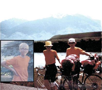
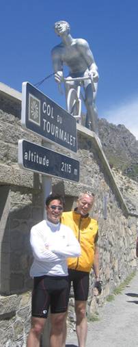
At the end of each day riding an average of 100 miles, we would look for some place to spend the night for free. We slept in schools, churches, private homes, and on two occasions in jail cells. It was an amazing experience and I still remember struggling up a mountain pass, looking up from my front wheel and seeing a massive male moose eating grass by the side of the road. I was hooked on cycling and during the next few years would spend summers riding across the U.S., touring England as well as doing some racing. I continue to enjoy cycling and in 2005 I had the pleasure of riding in the Pyrenees with my son Jason during the Tour de France (Figure 3).
Undergraduate Years
In 1973 I entered the University of Minnesota, Duluth with the intention of studying biology in preparation for medical school. During my first term I met two people who would have a lasting effect on my life and career. The first and most important encounter occurred in biology lab. Students worked together in groups of four. On the first day of class my group consisted of three freshmen and an irate sophomore. The sophomore (Tong Sun Thian), who was upset at having to defer to freshmen for choice of lab, would become my wife in 1978 (more about Tong Sun later).
The second important encounter was with Professor Conrad Firling, the biology professor who taught both the course and my lab section. Professor Firling delivered lectures in basic biology with enthusiasm and passion. I learned that he was willing to take undergraduates into his lab to work on projects in developmental biology. The first technique I learned was the proper way to wash glassware, and I washed a lot of glassware. I eventually progressed to bench work and helped Professor Firling develop an organ culture medium for Chironomous tentans, a model system for developmental biology. Tong Sun also joined the Firling lab and worked with me on this project.
As an undergraduate, I majored in biology and chemistry and benefited from excellent teachers and small classes. In addition to my work with Professor Firling, I worked on a summer project with Professor Robert Carlsen, an organic chemist. In spite of my growing interest in basic research, I applied to ten medical schools as well as a few graduate programs. I still envisioned a career in medicine and the graduate programs were a back-up option if I didn’t get into medical school. Of the medical schools to which I applied, Yale was by far the long shot. I was very surprised when I received the acceptance letter.
Yale University Medical School
My move to New Haven was a culture shock because of the inner city location and the academic environment. The medical school was located in a neighborhood troubled by crime and poverty. In school, I was intimidated by my classmates, many of whom came from Ivy League schools, some having had successful careers outside of medicine.
At Yale, all medical students were required to write a thesis based on original research, which we were to fit into our normal class schedule and vacations. My first research project involved dengue fever and allowed me to spend a summer working in a lab in Malaysia experiencing tropical epidemiological research. This gave me the opportunity to observe field research first hand, as well as perform bench work under more primitive conditions than I was used to at Yale. While this project was not successful, it was a great experience and led me to briefly consider tropical medicine as a career. For my thesis project I worked with Professor Denis Knudsen, a virologist in the department of epidemiology, studying the genetic diversity of rotavirus, a common cause of gastroenteritis in children.
Tong Sun and I married in 1978, after my first year in medical school. At Yale she worked in the Lab of Professor Caroline Slayman and obtained a masters degree in East Asian Studies in her spare time.
Clinical Training At Barnes Hospital
My career path following medical school was dictated early on by financial constraints. My medical school tuition was covered by a Public Health Service Corps scholarship that obligated me to work in a medically underserved community for three years following my residency training. As such, a career in research was not an immediate option. I decided on a residency in internal medicine and was assigned by the match program to a position at Barnes Hospital, which is affiliated with Washington University School of Medicine in St. Louis, Missouri. My career options changed during my final year at Barnes. Due to budget cuts, many of the Public Health Service programs lost funding. As a result, I was allowed to pay back my medical school scholarship by working in an academic hospital for three years, giving me the opportunity to explore basic research.
During my clinical training at Barnes Hospital, I became particularly interested in intensive care medicine. Patients admitted to one of the three intensive care units (medical, pulmonary and cardiac) were typically very unstable, requiring urgent intervention, often with medications acting on G protein coupled receptors (GPCRs) including adrenergic and muscarinic receptors to regulate their heart rate and blood pressure, as well as opioid receptors to control pain. My interest in intensive care medicine led me to apply for cardiology fellowships. I was particularly interested in the program at Duke University, which allowed fellows to work for several years in a basic research lab. Moreover, the laboratory of Robert Lefkowitz at Duke was doing pioneering research on adrenergic receptors, giving me the opportunity to explore basic research in an area relevant to cardiovascular and intensive care medicine. I was fortunate to be accepted into the fellowship program and the Lefkowitz lab.
Fellowship Training At Duke University
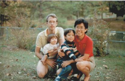
While in St. Louis, Tong Sun and I started our family. Our son Jason was born June 7, 1981, one week before I started my internship. Our daughter Megan was born November 28, 1983 during the last year of my residency. My only regret is that my training and call schedule prevented me from spending more time with my young family during this time. In June 1984 we packed up our belongings into a U-Haul trailer and I drove east to Durham, North Carolina with the family following by plane (Figure 4). We moved into a spacious but drafty two-story apartment a few miles from the University and I would often run to work, leaving the car for Tong Sun and the children. I had started running back and forth to work in St. Louis as my only way of getting exercise. The trip was approximately 5 miles one way, or 8 if I was working at the Veterans Hospital. I would carry my clothes in a backpack and shower at the hospital. My running commute at Duke was shorter, so I would often supplement that with a 5 mile run on the Duke track before going to the lab. During the first few years in the lab I was in the best physical shape of my life. Tong Sun had taken up running regularly in St. Louis, and in Durham we would often take the children to a local high school track and take turns running while Megan and Jason played in the long-jump sand pit or jumped on the cushions for the pole vault.
Duke University gave cardiology fellows the option of starting in a research lab before completing their 18 months of clinical rotations. This was perfect for me because I was tired of being on call every 2 to 4 nights and wanted to spend more time with my family. My first few weeks in the Lefkowitz lab were a bit awkward. As a senior resident in medicine at Barnes, I was in charge of a team of junior residents and medical students that cared for 20–30 patients. On joining the Lefkowitz lab I became the least experienced person in a group of very talented young scientists consisting of predominantly postdoctoral fellows and a few graduate students. I was not familiar with any of the techniques being used and had yet to familiarize myself with the literature leading up to work being done in the Lefkowitz lab at that time. My colleagues were very friendly, but also very busy with their own projects and I really didn’t know where to begin.
During the first months in the Lefkowitz lab, while learning basic techniques such as ligand binding and adenylyl cyclase assays, I became aware of the effort to obtain a cDNA clone of the β2 adrenergic receptor (β2AR). This was a collaborative project between the Lefkowitz group and Merck. Jeff Benovic, then a graduate student, had succeeded in purifying enough β2AR from hamster lung tissue to obtain several peptide sequences. These were being used to make degenerate and ‘guessed’ oligonucleotide probes to isolated clones from cDNA libraries. Richard Dixon, an experienced molecular biologist at Merck, was doing the cloning work. This was exactly the type of research project I hoped to be involved in, so I asked if I could contribute. At this stage of the project, almost all of the work was being done at Merck, Bob Lefkowitz decided that I could try to generate antibodies to purified β2AR, which could then be used to identify cDNA clones using expression cloning methods. The project was relatively simple. I would purify protein from hamster lung tissue and immunize rabbits. This kept me busy part time for several months and in my spare time I started testing various cell lines in an effort to find one that expressed high levels of β2AR for use in making cDNA libraries. I screened approximately 10 cell lines and found that many appeared to express relatively large amounts of β2AR. For a brief period of time I felt I was making a real contribution to the effort. Unfortunately, careful scrutiny of my data and methods by one of my experienced colleagues revealed that most of what I was detecting as β2AR was in fact non-specific binding of radioligands to membrane lipids. To this day I remember the feeling of disappointment and stupidity associated with these botched experiments. My antibody work was going a bit better and I was able to obtain antibodies that could recognize the receptor, but the polyclonal serum reacted with a number of other proteins on western blots and was not specific enough to be useful for cloning purposes.
In brief, my first five months in the lab were far from successful. Surprisingly, Bob allowed me to continue. Despite my having made no meaningful contribution to the cloning effort using the biochemical and pharmacological expertise in the Lefkowitz lab, Bob allowed me to spend time at Merck learning molecular biology from Richard Dixon. Over the next few months, I made several one-week trips to Richard’s lab at the King of Prussia site of Merck just north of Philadelphia. This amounted to intensive training sessions where I learned how to prepare and screen cDNA libraries. When I returned to Duke, Bob gave me his approval to set up a molecular biology lab within the Lefkowitz lab. Tong Sun, who had a masters degree in microbiology, was hired part time to help with our fledgling molecular biology effort and over the coming months I would be joined by a few other new postdoctoral fellows and a graduate student. Our initial goal was to screen libraries prepared by Richard Dixon. During this time I had experience with non-specific binding of another sort. Many of our initial “hits” turned out to be non-specific interactions with our probes. This led to many cycles of exhilaration and disappointment common to many research efforts.
After about a year of failure, we came to the conclusion that β2ARs were expressed at such low levels in most cells that we might not be able to isolate a clone from a cDNA library. Additionally, we had concerns about the degenerate and ‘guessed’ DNA probes we were using. Using degenerate probes meant that we had the correct sequence, but also many incorrect probes that could contribute to non-specific false positives. To avoid this, we took advantage of codon bias information to make a best guess of the coding sequence for several peptides. This would limit non-specific interactions; however, if we made too many errors in guessing the coding sequence, we had no chance of pulling out the clone. We realized that we might stand a better chance of pulling out a DNA fragment from a genomic library. Even if the genomic clone was incomplete and interrupted by introns, we reasoned that it would provide a larger, more specific probe that would allow us to make enriched cDNA libraries or identify rare clones in existing libraries. I made a genomic library from hamster lung DNA and sent an aliquot to Richard at Merck. The library was initially screened with a few long guessed probes at Merck and Duke, and both groups isolated a clone that surprisingly contained the full coding sequence with no introns. After almost two years of frustration, we had an amazing stroke of good fortune.
Cloning of the β2AR led to cloning of several other adrenergic receptor genes as well as an orphan receptor that turned out to be a member of the serotonin receptor family. The initial G protein coupled receptor (GPCR) clones revealed a common seven transmembrane architecture shared with rhodopsin, a GPCR specialized for the detection of light. It was subsequently determined that there were nine genes encoding distinct adrenergic receptor subtypes.
After having a few adrenergic receptor clones in hand, I began exploring approaches to understand receptor structure. I first took advantage of having closely related receptors that responded to adrenaline, but activated different signaling proteins and bound to specific synthetic agonists and antagonists. Chimeric receptors generated from the alpha and beta adrenergic receptors provided the first clues to the role of specific domains of the receptor in ligand binding and G protein coupling. However, these studies did not tell us how the receptor worked in molecular detail, and I started to think about obtaining a crystal structure. Several years earlier Deisenhofer and Huber had obtained the first crystal structure of a membrane protein, proving that membrane proteins could be crystallized and demonstrating the value of protein structure in understanding mechanisms. However, the photosynthetic reaction center was a naturally abundant protein that could be obtained from bacteria. In contrast, even in lung tissue, where the β2AR was most abundant, it represented a very small fraction of membrane proteins.
Stanford University
Early in 1989 I began considering career options. I was offered a junior faculty position at Duke, and interviewed for similar positions at Washington University in St. Louis, the University of California San Francisco and Stanford University. At Stanford, Professor Richard (Dick) Tsien, who had just moved from Yale, was building a new Department of Molecular and Cellular Physiology in a new building called the Beckman Center. I remembered Dick from his medical school lectures at Yale; he was one of our favorite teachers. I was impressed by his enthusiasm and vision for the new department at Stanford. Most of the faculty would be junior recruits and new to Stanford. I accepted a position and moved to Stanford shortly after the Loma Prieta earthquake in 1989.
The move to Stanford brought many challenges and opportunities. The greatest challenges were financial. We would be moving from one of the most affordable housing markets to one of the most expensive. While I would be earning more at Stanford, our mortgage would triple and we would have only one income, as Tong Sun had started medical school. To accommodate the larger mortgage and medical school tuition, I took a 48 hour shift as an emergency room physician at a nearby hospital once or twice a month. During the six years at Stanford, with Tong Sun a full time medical student, I had the opportunity to spend more time with Jason and Megan as their primary care giver, making up for some of the time I missed during my residency training.
In the lab I focused on two questions: understanding the structure and mechanism of activation of the β2AR, and determining the physiologic role of specific adrenergic receptor subtypes. Cloning and pharmacological studies had identified 9 adrenergic receptor subtypes coded by 9 different genes: three βARs, three α1ARs, and three α2ARs. The drugs available at that time were not sufficiently selective to allow assignment of specific functions to each receptor subtype. To address this problem we used recently developed methods to disrupt genes in mice. In collaboration with my Stanford colleague Greg Barsh, we created strains of knockout mice for 5 of the 9 adrenergic receptor genes (β1AR, β2AR, α2AAR, α2BAR and α2CAR) and were able to assign their roles in cardiovascular function and behavior.
At the same time we were laying the foundation for future biophysical and crystallography studies. We continued to use mutagenesis and chimeric receptors to provide a more detailed map of the functional domains of adrenergic receptors. We were also investigating receptor biosynthesis and exploring protein engineering techniques that would enable expression and purification of sufficient quantities of receptor for crystallography. My own efforts in the lab focused on comparing different expression systems (mammalian cells, insect cells, bacteria and yeast), and establishing an efficient purification protocol. By 1993 we were able to express and purify sufficient quantities of functional β2AR in insect cells to begin using fluorescence spectroscopy, one of the most sensitive biophysical techniques, to investigate receptor structure. We labeled purified β2AR with small, environmentally sensitive fluorescent probes, most often attaching them to a single reactive cysteine introduced into a specific domain. Using this approach we were able to observe ligand-induced conformational changes in real time. These relatively simple fluorescence experiments provided important insights into the dynamic character of the β2AR that would ultimately guide our strategies for crystallizing the β2AR and the β2AR-Gs complex.
With incremental improvements in expression and purification strategies we were able to produce enough β2AR to start crystallography trials. My Stanford colleague Bill Weis taught me the basics of setting up crystallography trials and would continue to be my crystallography mentor. These initial trials introduced me to the many types non-protein crystals (salts, detergent, lipids) that one encounters trying to crystallize membrane proteins. Each cycle of failed trials led to a reassessment of the approach followed by modifications to the strategy and a new round of trials. The increasing number of prokaryotic membrane protein structures being published during this time provided a constant source of inspiration. But it wasn’t until 2004 that we obtained the first crystals of the β2AR. These crystals were very small (β2AR crystals to the ESRF. Using a high intensity 5 micron beam we were able to see diffraction compatible with a protein crystal at a resolution of approximately 20Å. While we were disappointed in the poor quality of the diffraction, we were encouraged by the fact that we were able to form crystals of the β2AR. This was an important milestone in the effort and suggested that a crystal structure of the β2AR was not an impossible goal. With this milestone, I felt I could begin to involve postdoctoral fellows in the effort.
In 2005 Dan Rosenbaum and Søren Rasmussen, two very talented and intrepid postdoctoral fellows, joined the lab with the goal of crystallizing the β2AR. Søren and Dan took two different approaches to generate better quality crystals of the β2AR. Søren identified antibodies that bound to a particularly flexible region of the receptor and Dan used protein engineering to replace the same region of the β2AR with T4 lysozyme (T4L), a highly crystallizable soluble protein. Both approaches were designed to minimize conformational flexibility and increase the amount of polar surface area for forming crystal lattice contacts. During 2006 we obtained crystals using both approaches combined with a newly developed lipid-based media known as bicelles consisting of a mixture of lipid and detergent. Initial crystals of the β2AR-Fab and the β2AR-T4L fusion protein complex both diffracted to below 4 Å. We subsequently obtained a 3.4Å structure of the β2AR-Fab complex grown in bicelles. This was our first look at the three dimensional structure of the β2AR, but a higher resolution structure would soon follow.
In the fall of 2006 we sent purified β2AR-T4L complex to Vadim Cherezov in the lab of Raymond Stevens at Scripps. Vadim had trained with Martin Caffrey at the Ohio State University. Martin’s lab had recently developed miniaturized, high-throughput methods for lipidic cubic phase (LCP) crystallography. We previously explored the use of LCP methods to crystallize the β2AR in 1999 in collaboration with Peter Nollert; however, at that time the methods were very labor intensive and used relatively large amounts of protein to screen very few conditions. The methods developed in Martin’s lab together with the robot built by his team enabled screening of thousands of conditions with a few milligrams of protein. Vadim had recently joined the Stevens lab, bringing with him a LCP robot on loan from Martin Caffrey. This collaboration led to a 2.4 Å structure of the β2AR-T4L complex. The fusion protein strategy developed for the β2AR has since been successfully applied to a growing number of other GPCRs.
These first β2AR structures represented inactive states; however, our goal had been to understand the mechanism by which agonist binding leads to G protein activation. At a Gordon Conference in 2005, I met Roger Sunahara, a kindred spirit who had crystallized the G protein Gs during his postdoctoral fellowship in the lab of Al Gilman. Roger and I soon became friends and started a very enjoyable and fruitful collaboration. During the next 5 years our labs would use a variety of methods to study activation of the Gs by the β2AR and together with an incredible network of colleagues (outlined in my Nobel Lecture) we would accumulate the reagents and expertise to stabilize and crystallize the β2AR-Gs complex. The β2AR-Gs crystal structure was published in 2011 together with two companion studies using single particle electron microscopy and deuterium exchange mass spectrometry to characterize the dynamic aspects of this complex. These combined studies provided unprecedented insights into GPCR signaling at a molecular level.
As noted above, I have benefited from mentorship, advice and collaborations with colleagues from a broad spectrum of disciplines. I have been particularly privileged that my wife Tong Sun Kobilka has worked with me in the lab on and off for more than 30 years providing emotional, intellectual and technical support. It has been great to share the joy of scientific discovery with her and to have her encouragement when research wasn’t going well or funding was tight. My career in basic research has been very rewarding from several perspectives: discovering new knowledge, working with many brilliant and often intimidating students and postdoctoral fellows, developing friendships with scientists throughout the world, and having the flexibility to spend time with my family. I am very grateful to be honored with a Nobel Prize in Chemistry for my work on G protein coupled receptor, and for the recognition it brings to my colleagues in the field.
This autobiography/biography was written at the time of the award and later published in the book series Les Prix Nobel/ Nobel Lectures/The Nobel Prizes. The information is sometimes updated with an addendum submitted by the Laureate.
Nobel Prizes and laureates
Six prizes were awarded for achievements that have conferred the greatest benefit to humankind. The 12 laureates' work and discoveries range from proteins' structures and machine learning to fighting for a world free of nuclear weapons.
See them all presented here.

