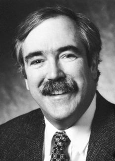
Eric F. Wieschaus
Biographical

I was born in South Bend, Indiana on June 8, 1947, one of that large bumper crop of babies born in the United States after World War II. My family moved to Birmingham Alabama in 1953 when I was six. Although Birmingham was already a major industrial center in the South, the city still had the small town character of most Southern cities at the time. My brother, my three sisters and I could go exploring in the woods near our house, and collect frogs, turtles and crayfish from the local streams and lake. I went to Catholic grade schools and, when I was fourteen, took a 6:45 bus every morning across the city to make it to the only Catholic high school by 8:30. Though I did well in my science and math courses, I did not see myself in a career in science. I played piano and read books, but spent most of my time painting and drawing pictures. I dreamed of becoming an artist when I grew up.
In the summer between my junior and senior years, I went to Lawrence, Kansas, to a program funded by the National Science Foundation to encourage high school kids to become scientists. For the first time in my life I was with kids who were smarter than I, who cared about science, and who talked about books and art. I felt as though I had finally found a group to which I belonged; in these surroundings I was able to conquer the shyness and insecurity that plagued me in my own high school back in Birmingham. In the laboratory associated with the Zoology course, I dissected animals for the first time, from fish up the vertebrate ladder to fetal pigs. I was invited back the following summer to work in the neurobiology lab of Nancy and Dennis Dahl. My work involved more dissection, this time removing vagus nerves from large land tortoises, stripping off the outer sheaths and recording the electrical depolarization when they were stimulated. The Dahls’ generosity in opening their lab to a high school senior still amazes me. I can’t believe I produced much useful data, but the experience was enough to convince me that I wanted to become a scientist. By the time I started college at Notre Dame, there was no doubt in my mind that I would major in biology.
In my sophomore year at Notre Dame, I needed money and found a job preparing fly food in a Drosophila laboratory run by Professor Harvey Bender. In Bender’s lab, I encountered my first fruit flies and learned basic genetics. Though I liked working in a lab, genetics did not excite me as much as the embryology courses I was then taking from Kenyon Tweedel. Tweedel seemed to have a continuous supply of living embryos from a variety of different species. I will never forget the thrill of seeing cleavage and gastrulation for the first time in living frog embryos. I immediately wanted to understand why cells in particular regions of the developing embryo behaved the way they did. What were the mechanisms that made them different from each other? What forces drove such dramatic rearrangements in the cytoplasm and the shape of cells?
In my last years at Notre Dame, I became increasingly active in the student effort against the war in Vietnam. I collected petitions, joined in protest demonstrations and applied for conscientious objector status to avoid being sent to Vietnam. It was, however, very unlikely that my local draft board would grant me such status, given that I had not been raised in one of the traditionally pacifist religions. In spite of my somewhat uncertain future, I decided to begin graduate school in biology and was accepted to Yale University. Bender was worried about my draft status and wrote to Donald Poulson, the only Drosophila geneticist he knew at Yale, telling him about my problems and asking him to look after me while I was in New Haven. When I arrived in New Haven, Poulson had a place set up for me in his lab. He was so very kind that I didn’t have the courage to tell him that after three years of washing fly bottles at Notre Dame, I never wanted to see another fly, much less work on flies for my thesis.
In the 30’s and early 40’s, Poulson had described the basic embryology of Drosophila and had characterized one of the first mutants with an interesting interpretable phenotype during embryonic development (the neural defects associated with deletions of the Notch locus). Until that point, I had thought all developmental genetics of flies involved eye colors and bristles and other aspects of adult morphology. It had never occurred to me that flies had embryos, or that Drosophila embryogenesis was characterized by the same kinds of spectacular cell movements seen in the classically studied embryos of vertebrates. I learned all that from Poulson.
In my second year in graduate school, I switched to Walter Gehring’s lab to learn in vivo techniques for culturing embryos. Gehring had just set up his lab in the medical school, so it was still very small, much smaller than what it was to become after his return to Basel two years later. Because I was his only student, working directly with him was a wonderful opportunity to learn how experimental science is done. For my first experiments in his lab, I set out to investigate whether cells at the blastoderm stage were already determined to form specific discs. My plan was to remove single cells from defined regions of the blastoderm and culture them in adult abdomens surrounded by genetically marked “feeder cells.” My last years in New Haven and almost all my time in Basel were spent dissociating embryos and trying different culturing procedures, but I was never able to get single isolated cells to survive. Fortunately, at some point along the way, I had decided I needed to know what normal cells did in embryos that weren’t homogenized or subjected to my in vivo culturing techniques. It was those experiments, initially planned as controls for my more ambitious cultures, that eventually constituted my thesis. I used X-ray induced mitotic recombination to mark clones derived from single cells. In contrast to the restricted clones produced by irradiation of larvae, such clones extended between the wing and leg of the adult fly, indicating that the blastoderm cell that gave rise to the clone could not yet have been determined with respect to either disc. On the other hand I could never find clones that overlapped adjacent legs. Because legs were derived from different segments, my results suggested that if blastoderm cells were determined for anything, they were determined for segments rather than discs.
In my last year in Basel I started a collaboration with Elisha Van Deusen and Larry Marsh using pole cell transplantation to make genetically mosaic ovaries. We wanted to use such mosaics to determine whether particular maternal effect mutants block gene activities in the germ cells themselves, or whether they identified genes that were active in the overlying follicle cells. Although most of the mutants we tested did not have interesting phenotypes, there was one, fs(1)k10, that caused an abnormal pattern in the egg shell. Since the shell is secreted by the follicle cells during oogenesis, we expected the defect to depend on the genotype of those cells. To our surprise, the mutant was germ line dependent. Those transplants provided the first evidence for an organizing principle that emanated from germ cells and controlled patterning of the overlying follicle cells. The embryos that developed in k10 eggs were also abnormal, but in a way that I did not understand at the time. It took Christiane Nüsslein-Volhards‘s work on dorsal for me to re-interpret it in terms of dorsal ventral polarity.
I met Christiane (Janni) Nüsslein-Volhard two months before I left Basel to begin my postdoctoral work with Rolf Nöthiger in Zurich. Janni had come to Basel to learn Drosophila embryology and we thus had many interests in common. Even after I had left for my postdoctoral work in Zurich, I would come back to Basel, in part to finish experiments, but also always to have dinner with her. We would talk science and plan experiments we eventually wanted to do together.
In much of my work in Zurich, I continued to use the cell lineage techniques of my thesis work, but now to analyze the development of sexually dimorphic structures. Janos Szabad and I developed efficient procedures for making germ line mosaics using K10 and mitotic recombination. In collaboration with Trudi Schüpbach, we also studied the cell lineage of the embryonic epidermis. Those studies paralleled a similar analysis that Janni Nüsslein-Volhard had begun with Margit Lohs using laser ablation. Both studies suggested that segmental units might be established as three to four cell wide stripes at the blastoderm stage. By far, however the most important thing that happened to me at Zurich was my deepening relationship with Trudi Schüpbach, who became my close friend and occasional scientific collaborator, but also an enormous emotional support throughout my life. We eventually married, in Princeton in 1983. Life with her, and with our three daughters Ingrid, Eleanor and Laura, has kept me busy and provided a needed balance to the demands of the lab.
In 1978, I moved on to my first independent job, at the European Molecular Biology Laboratory in Heidelberg. The lab had just been built and was intended as an international meeting ground for scientists from the various member nations. Although there were no permanent contracts at the time, the position as group leader gave me independence to pursue my interest in embryos, without major teaching obligations or being required to explain every step in my experimental plans to get the necessary funding. The job was as extraordinary opportunity, one that I regret is not given to more young scientists at the beginning of their careers. The most attractive feature of the move to Heidelberg, however, was that the EMBL had also offered a similar position to Janni. This gave us the chance to realize many of the experiments, and test many of the speculations developed over the long dinner conversations in Basel.
Although we tried to keep our own individual research projects going, most of our time at the EMBL was spent on the joint mutagenesis experiment. Because handling large numbers of flies was essential if we were going to saturate the fly genome for mutations affecting embryonic development, we first had to test genetic selections to kill off specific genotypes at particular generations, and establish techniques for making large numbers of microscope preparations. We also continued to discuss developmental issues, recent models for patterning in embryos and, as the mutagenesis screens got underway, the interpretation of the particular defects observed in the various mutant lines. Those years were probable the most exciting, intellectually stimulating ones of my entire scientific career. A special feature of those mutagenesis experiments was that almost every day we could expect to encounter a new phenotype, a phenotype that would force us to re-evaluate some long held assumption about embryonic development.
I moved from Heidelberg to Princeton in 1981. Since then I have taught genetics and development courses at the graduate and undergraduate level. The Heidelberg experiments continue to provide a rich source of inspiration for further research. After arriving in Princeton, Trudi Schüpbach and I carried out similar large scale mutagenesis screens for maternal effect mutants. The loci identified in those screens, as well as in a comparable screen made by Janni’s lab in Tübingen, allowed Drosophila oogenesis to come to rival embrogenesis as a ideal system for studying patterning. I have also continued to be interested in segmentation genes, as well as genes affecting segmental identity. Peter Gergen, Jym Mohler and Doug Coulter began their analyses of runt, hedgehog and oddskipped in my lab at Princeton and the extradenticle gene was analysed by Mark Peifer and Cordelia Rauskolb. Our work on the armadillo was started by Bob Riggleman and Paul Schedl, and was continued by Mark Peifer.
Much of my current work centers on genes controlling cell shape changes during gastrulation (with Sue Zusman, Suki Parks, Dari Sweeton, Mike Costa), and genes for the establishment of the early cytoskeleton (work with Lesilee Rose, Eyal Schejter, Marya Postner). The mutagenesis experiments in Heidelberg were less successful in identifying genes directly involved in such specific morphological changes. We have consequently designed alternate genetic procedures involving translocations to identity such genes and have initiated an analysis of their roles within the cell. Overall the Princeton years have seen an increasingly cell biological turn to my research. My work has always had a strong visual component (probably to assuage my suppressed teenage desires to be an artist or painter). What I did not realize until late in my development as a scientist is that morphology and cell biology are actually the same scientific areas, or at least that the latter provides the molecular explanation of the former.
This autobiography/biography was written at the time of the award and later published in the book series Les Prix Nobel/ Nobel Lectures/The Nobel Prizes. The information is sometimes updated with an addendum submitted by the Laureate.
Nobel Prizes and laureates
Six prizes were awarded for achievements that have conferred the greatest benefit to humankind. The 12 laureates' work and discoveries range from proteins' structures and machine learning to fighting for a world free of nuclear weapons.
See them all presented here.
