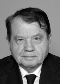
Luc Montagnier
Biographical

I was born on August 18, 1932 in Chabris, a “bourg”, larger than a village but smaller than a town, located in Berry south of the Loire Valley. This was – and still is – a region of agriculture with some renowned products such as welsh rabbit, goat cheeses and white asparagus. It was the place where my mother had grown up but, in fact, I never lived there.
On my father’s side, his parents came from Auvergne, a province in the centre of France, made of rich plains and old volcanoes, the latter probably being at the origin of my family name: Montagnier, the man living in mountains.
In his youth, my father had caught a terrible disease: streptococcal arthritis, ending in irreversible lesions in the aortic valves. He was therefore declared unfit for military service and had to find a sedentary job: he became an accountant and excelled in this profession, which implied, at that time, mainly hand-written work. He started working in the Poitiers area and then moved a little farther north to Châtellerault, a small city between Tours and Poitiers.
As an only child, I was cherished by my mother, a housewife, but two events dominated this pre-war period, of which I keep a vivid memory:
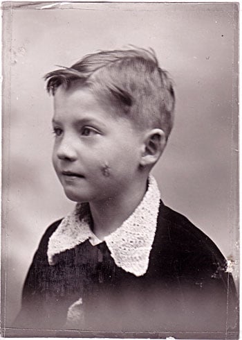
I was badly injured by a high speed car while crossing a main road: multiple wounds of which I keep some visible scars. After two days in a coma, I emerged as if I was born again, at the age of 5 (Figure 1).

… and two years later came the declaration of war in 1939, while the whole family was harvesting grapes in the vineyards of my mother’s brother. I still remember the images in a newspaper of Warsaw ruins after a bombing by German planes.
And then, in 1940, came the “real” war: the German invasion, my parents and I leaving their house (close to a risky railway station), fleeing on the roads in a little car, and finally more exposed to German bombing during this “exodus” than if we had stayed home.
The first year of German occupation was terrible, in that we had no food reserves and most of the time we were starving. I was a rather puny boy and during the four years of the war did not gain a gram! The “ersatz” did not stimulate my appetite, when I was dreaming of chocolate and oranges! My father had chronic enterocolitis and, worse, my grandfather (his father) was diagnosed with rectal cancer. He died in 1947 after terrible suffering and each time I visited him, I could see the inexorable progression of the disease. This affected me so much that it is probably one reason why I decided later to study medicine and to start research on cancer.
In June 1944, our house (so close to the railway) was partly destroyed − this time by an Allied bombing. I keep a mixed feeling of this year of the liberation of France. It was a great relief but I could not forget also the vision of so many dead people, civilians and soldiers, and the images of skinny deportees released from concentration camps. I will hate wars and their atrocities for the rest of my life.
At high school I did well, being usually ahead of my classmates. This is when I became curious about scientific knowledge, having left behind my religious Catholic belief.
Following the example of my father, who was tinkering in his leisure days with electric batteries, I set up a chemistry laboratory in the cellar of the new house which was requisitioned to accommodate us. There, I enthusiastically produced hydrogen gas, sweet-smelling aldehydes and nitro compounds (not nitro-glycerine!) that had the unfortunate habit of blowing up in my face.
I was delighted to read – in popularised books – the impressive progress of physics, especially atomic physics. Being good in physics and chemistry – but not as good in maths – I decided not to prepare to compete for the “Grandes Ecoles” but instead to register both at the School of Medicine and the Faculty of Sciences in Poitiers. My goal was in fact to start a research carrier in human biology, but there was no such specialty in Poitiers, either in Medicine or in Sciences. Since both the Faculty and School were within walking distance, I could spend the morning at the hospital and the afternoon attending courses in botany, zoology and geology, which were the main disciplines of the degree course in Sciences.
Fortunately the new Professor of Botany, Pierre Gavaudan, was a very atypical professor in that his scientific interests went far beyond the classification of plants. In fact, I owe him for having opened me a large window on what was the beginning of a new Biology, the DNA double helix, the in vitro synthesis of proteins by ribosomes and the structure of viruses.
At the same time, I was installing at home a device combining a time-lapse movie camera and a microscope, thanks to a gift by my father. This allowed me to do my first research work. I was studying a phenomenon known since 1908 as the phototaxy of chloroplasts: the property of some algae living at the surface of ponds to orient their large unique chloroplast according to the intensity of light; if the light was too intense, the chloroplast turned inside the tubular cell to present its edge. In dark or weaker light, the chloroplast, a flat plate, exposed its larger surface. The phenomenon took a few minutes, which could be analysed by time-lapse cinematography. Using different glass filters, I could show that it was not the wavelength absorbed by the chlorophyll (red light) which regulated the orientation of the chloroplasts but indirectly some yellowish pigments absorbing the blue light. I was very proud, at the age of 21, to defend this work as a small thesis at the Faculty of Sciences of Poitiers. I was asked by my mentor, Pierre Gavaudan, to do research also on a literature-based subject: the L-forms of bacteria. This allowed me to make my first incursion – not the last – into the world of filtering bacteria. I could only find the references on this controversial subject at the library of the Institut Pasteur in Paris. This was indeed the time when I left Poitiers for Paris, where I was able to complete my medical studies as well as explore some aspects of biology closer to human beings, particularly neurophysiology, virology and oncology.
Having been hired as an assistant at the Sorbonne at the age of 23, I started learning old-fashioned technologies derived from Alexis Carrel‘s work on chick embryo heart cultures, as well as that of human cell lines in monolayers. Although my research was not productive at all, I keep from this period a solid expertise of Pasteurian technologies for working in perfectly sterile conditions without the use of antibiotics.
In 1957, the first description of infectious viral RNA from the tobacco mosaic virus by Fraenkel-Conrat and Gierer and Schramm determined my vocation: to become a virologist using the modern approach of molecular biology.
I started with the foot and mouth virus and then, in Kingsley Sanders’ laboratory at Carshalton near London, I was proud to identify for the first time an infectious double-stranded RNA from cells infected with the murine encephalomyocarditis virus, a small single-stranded RNA virus. This demonstrated for the first time that RNA could replicate like DNA by making a base-paired complementary strand.
In order to perfect my knowledge of oncogenic viruses, I moved from Carshalton to Glasgow where a new Institute of Virology had been recently inaugurated, headed by a remarkable virologist, Michael Stocker, and where many high-ranking visitors, among them Renato Dulbecco, were spending sabbatical years.
Working on a small oncogenic DNA virus, polyoma, I could show there, with I. Macpherson, a new property of transformed cells, that of growing in soft agar. Using this technique, it was easy to detect the transforming capacity of polyoma virus and its DNA. We showed that naked DNA alone carried all the oncogenic potential of the virus. This now looks pretty obvious, but it was not so at that time.
Back to France at the Institut Curie, I extended this finding to a number of cancer cells, transformed or not by oncogenic RNA or DNA viruses. However, this property allowed me to distinguish some in vitro steps in the process of transformation, which were correlated with some modifications of the plasma membrane and of the carbohydrate layer surrounding it.
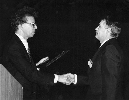
A great mystery remained at that time: that of the replication of the oncogenic RNA viruses, now known as retroviruses. Howard Temin (Figure 2) had proposed the hypothesis of a DNA intermediate, but other possibilities could be considered. I myself tried to find a double-stranded RNA specific of the Rous sarcoma virus, a virus able to infect and transform chick embryo cells. I indeed isolated double-stranded RNA sequences, but they were of cellular origin and existed at the same level in non-infected cells! With Louise Harel, I later showed that this RNA was partly coming from repetitious sequences of DNA. In retrospect, it could at least in part represent the recently identified interfering RNAs involved in the negative control of messenger RNA translation.

In 1969–70, the isolation of an RNA-polymerase associated with the viral particles of the vesicular stomatitis virus led to the idea that perhaps a key enzyme was also associated with the oncogenic RNA viruses. Indeed, Howard Temin and Mizutani, and independently David Baltimore, discovered in 1970 a specific enzyme associated with Rous sarcoma virus (RSV), the reverse transcriptase (RT), capable of reversely transcribing the viral RNA into DNA.
At about the same time, Hill and Hillova in Villejuif, France, demonstrated that the DNA extracted from RSV transformed cells was infectious and carry the genetic information of the viral RNA, confirming that the enzyme was working faithfully in infected cells.
I myself, with P. Vigier, confirmed and extended this discovery by showing that the infectious DNA was associated with the chromosomal DNA of the cells, showing integration of the proviral DNA, as earlier postulated by Temin.
Work on the chicken RSV was extended to similar viruses in mammals, so that many researchers at that time believed that RT activity was a new, highly sensitive tool for detecting similar viruses in human leukaemia and cancer. This was stimulated by the generously funded virus-cancer program launched by America’s National Institutes of Health. Unfortunately, the hunt for human retroviruses was basically unsuccessful but led to important basic work on the molecular biology of animal retroviruses.
In 1972, I was asked by Jacques Monod, then head of the Institut Pasteur, to create a research unit in the newly created Department of Virology of the Institute. I accepted, and this new laboratory allowed me to develop new avenues of research within the general theme of Viral Oncology, the ultimate goal remaining the detection of viruses involved in human cancers.
Thus, I became interested in the mechanism of action of interferon and its role in its expression of retroviruses. I came into this field after having demonstrated the biological activity of interferon messenger RNA in collaboration with two world-renowned experts in the field, Edward and Jacqueline De Maeyer.
From 1973 on, Ara Hovanessian and his co-workers joined my unit and brought a new dimension: the complex biochemical mechanism sustaining the antiviral activity of this remarkable group of cellular proteins.
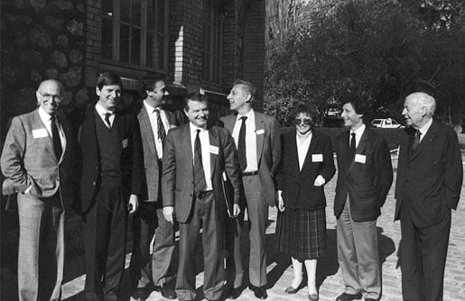
In 1975, two other researchers joined my unit and brought their expertise on murine retroviruses: J. C. Chermann and his collaborator, Françoise Barré-Sinoussi (Figure 3). The latter mastered particularly the detection of retroviruses by their RT activity. I convinced them to participate in a joint study inside the unit to look again for retroviruses in human cancers. We started in 1977 with blood samples coming from different Paris hospitals and biopsy specimens.

Two advances made in other laboratories boosted this search:
In Villejuif, France, Ion Gresser had prepared a potent antiserum neutralising any molecule of alpha endogenous interferon produced by individual cells. This interferon, we realised, was produced by mouse cells induced to express some of their endogenous retroviruses. Its blockade by the antiserum increased by up to 50 times the production of endogenous retroviruses in the culture medium. We could conclude that, despite the fact that endogenous retroviruses have been integrated in the genome of vertebrates for millions of years, their expression is still controlled by the interferon system, like that of exogenous viruses.
At about the same period, the discovery by Denis Morgan and Frank Ruscetti in Dr. Gallo’s laboratory of a growth factor allowing the in vitro multiplication of human T lymphocytes (TCGF, then named interleukin 2, Il2) made it possible to propagate T lymphocytes in sustained cultures.
We knew at that time that some retroviruses involved in mouse mammary tumour formation (MMTV) could not only be expressed in the tumour cells but also in the circulating lymphocytes.
Taking advantage of these two advances, we started a search for retroviruses in human cancers. Using anti-interferon serum and Il2, we focused particularly on the T lymphocyte cultures from breast cancer patients.
Indeed, in 1980, we were able to detect a DNA sequence close to that MMTV, not only in the cells of an inflammatory breast cancer (from a North African woman), but also in her cultured T lymphocytes. A second patient showed similar results.
Unfortunately, the molecular tools we had at that time could not tell us whether we were dealing with endogenous retroviral sequences or with an exogenous virus. Nowadays, having access to more powerful technologies, I am planning to reinitiate these studies.
But in 1983, the same approach, the use of anti-interferon serum, and the use of long term cultures of T lymphocytes greatly facilitated the isolation of HIV.
My involvement in AIDS began in 1982, when the information circulated that a transmissible agent – possibly a virus – could be at the origin of this new mysterious disease. At that time there were only a few cases in France, but they attracted the interest of a group of young clinicians and immunologists. They were looking for virologists, especially retro-virologists, as a likely hypothesis was that HTLV – the only human retrovirus known so far, recently described by R. C. Gallo – could be involved. Retrovirus causing leukaemia in rodents often also causes a wasting syndrome, which could be the result of secondary immune depression. This was also the case of patients suffering from leukaemia induced by HTLV.
A member of the working group, Françoise Brun-Vézinet, was a former student of the virology course that I was then directing. She called me up to organise the search for the putative retrovirus from a patient presenting with an early sign of the disease, lymphodenopathy. The patient was a young gay man who had been travelling to the USA and who was consulting Dr. Willy Rozenbaum – one of the leaders of the working group – for a swollen lymph node in the neck.
The reasoning was that if we were to find a virus at this early stage of the disease, it could be more a cause than a consequence of the immune depression.
Another incentive to start this research was a request from the producers of hepatitis B virus vaccine in the industrial subsidiary of the Institut Pasteur. They were using plasmas from American blood donors and were concerned by the risk of transmission of the AIDS agent through their procedure of viral antigen purification.
The lymph node biopsy arrived on January 3, 1983, a date which I remember well because it was also the first day of the virology course at the Institut Pasteur, which I had to introduce. I could only dissect the small hard piece at the end of the day. I dissociated the lymphocytes with a Dounce glass homogeniser and started their stimulation in culture with a bacterial mitogen, Protein A, known as an activator of B and T lymphocytes, since I did not know which fraction of lymphocytes could produce the putative virus. Three days later, I added the T cell growth factor I had obtained from a colleague working in the laboratory of Jean Dausset.
The T cells grew well. As previously established in a protocol for the search of retrovirus in human cancers, it was decided with my associates, Françoise Barré-Sinoussi and Jean-Claude Chermann, to measure the RT activity in the culture medium every 3 days. On day 15, Françoise showed me a hint of positivity (incorporation of radioactive thymidine in polymeric DNA), which was confirmed the following week.
We had evidence of a retrovirus, but this was just the beginning of a series of questions:
• Was it close to HTLV or not?
• Was it a passenger virus or, on the contrary, the real cause of the disease?
In order to answer these basic questions, we had to characterise the virus biochemically and immunologically, and to do that, we needed to propagate it in sufficient amounts. Fortunately, the virus could be easily propagated on activated T lymphocytes from adult blood donors. No cytopathic effect was observed with this first isolate, but unlike HTLV infected cultures, no transformed immortalised cell lines could emerge from the cultures, which always died after 3–4 weeks as do normal lymphocytes.
By contrast, subsequent isolates I made from culture of lymphocytes of sick patients with AIDS were cytopathic for T lymphocytes culture and – we discovered later – could be cultivated in larger amounts in tumour cell lines derived from leukaemia.
Shortly after the virus isolation, my co-workers and I were able to show that it was not immunologically related to HTLV, and in electron microscopy, it was very different from HTLV viral particles. In fact, as soon as June 1983, I noticed the quasi-identity of our virus with the published electron microscopy pictures of the visna virus in sheep, the infectious anaemia virus in horses and the bovine lymphocytic virus: it was a retrolentivirus, a sub-family of viruses causing long-lasting disease in animals without immunodeficiency.
This indicated clearly that we were dealing with a virus very different from HTLV, and my task was now to organise a team of researchers to accumulate evidence that this new virus was indeed the cause of AIDS.
It was an exciting period, since every Saturday morning when we had a meeting in my office, new data were brought by my associates favouring the causative role of the virus. The viral isolates were called LAV, for Lymphadenopathy Associated Virus, when it was isolated from patients displaying swollen lymph nodes, a frequent sign of the early phase of infection. The isolates made from patients with full-blown AIDS were called Immuno Deficiency Associated Viruses (IDAV). The latter generally grew better in T lymphocyte culture and induced the formation of large syncitia, resulting from the fusion between several infected cells. Some of them – we found out later – could also multiply in continuous cell lines of B or T cell origin. The latter property greatly facilitated the mass production of the virus for commercial use.
By September 1983, I was able to make a synthesised presentation of all our data favouring a causal link between the virus and the disease at a meeting on the HTLV organised by L. Gross and R. Gallo at Cold Spring Harbor.
This presentation was received with scepticism by a small audience (it was a late night session) and the HTLV theory still prevailed. Mentally, most attendants were not prepared to accept the idea of a second family of retroviruses (lentiretroviruses) existing in humans and causing immune deficiency, and having no counterpart in animals!
This situation is not infrequent in science, since new discoveries often raise controversy. The only problem is that it was a matter of life and death for blood transfused people and haemophiliacs, since a serologic blood test using our virus antigen was already working at laboratory scale but awaited industrial and commercial development.
This came in 1985, after two other teams of researchers, first that of Dr. Gallo at the NIH in early 1984 and that of Jay Levy in San Francisco, confirmed and extended our findings. In particular, Dr. Gallo and his associates gave more strength to the correlation between the virus and the disease, improved the detection of the antibody response and were able to grow several viral strains, including ours, in T cell lines of cancer origin. Meanwhile, my co-workers showed the tropism of the virus for CD4T cells and identified the CD4 surface molecule as the main receptor to the virus.
The rest of the story is described in the next chapter. I would just like to illustrate how I discovered what I believe are two important phenomena for explaining the destruction of the immune system induced by HIV.
During the latent phase of the infection, no virus is found in the blood. It is mostly localised in lymphocytes of lymphatic tissues and yet, we found that most of the lymphocytes present in the blood are sick! In 1987, a young visitor from Sweden, Jan Alberts, came to my lab. He wanted to cultivate human lymphocytes in a serum-free synthetic medium and to learn some technologies about HIV culture. The surprise came when we compared the viability in his medium of lymphocytes from healthy donors and those from HIV infected patients, even in their early asymptomatic stage of infection. While the former could survive several days without dying, the majority (more than 50%) of the latter died very quickly. Addition of interleukin 2 partially prevented their death.
When we used normal culture medium supplemented with foetal calf serum, the same difference was observed, although the survival time of the lymphocytes from infected patients was longer.
It did not take very long before three of my collaborators found the reason for such deaths: apoptosis. This is an active process by which the cell “decides” to die in a clean way, without releasing too many toxic compounds into the medium.
It is a physiological way of preventing abnormal proliferation of activated lymphocyte clones, but here the phenomenon was enormous and bore not only on the main cellular target of HIV infection, CD4+ T-lymphocytes, but also on cells which were not infectable by the virus, such as CD8+ T-lymphocytes, B-lymphocytes, monocytes, natural killer cells … Clearly, it was a general phenomenon, the culture simply revealing a predisposition to apoptosis of the majority of circulating blood cells, although most of them were not infected. Indeed, my collaborator Marie-Lise Gougeon found a very good relation between in vitro apoptosis and the in vivo observed drop of CD4 T cells in patients.
We have spent a lot of time trying to find the origin of this massive apoptosis, without finding a completely satisfactory explanation: the most likely is the intensive oxidative stress existing in patients since the beginning of their infection. This is also a finding I am very proud of: although oxidative stress has been – and still is – completely overlooked by AIDS researchers, it is likely to aggravate the wrong activation of the immune system at the origin of its decline and also it triggers inflammation through the production of cytokines.
Of course, the next question arises: what are the factors causing oxidative stress: viral proteins, fragments of viral DNA, co-infection with mycoplasmas? Even after 25 years, we still do not know the complete answer. But the phenomenon does exist and needs to be treated, while most AIDS clinicians do not care about it at all!
The treatment by combined antiretroviral therapy has, without doubt, changed the prognosis of this lethal disease, from a death sentence to an almost “normal” life. However, the virus is still there, ready to multiply when the treatment is interrupted, and not all HIV infected patients in the developing world have access to it. And the epidemics still kill 2–3 million people each year. It is thus absolutely necessary to resolve these problems. Basic research, as well as clinical research, has to be continued.
In addition, I realised in the 1990s that research should not only be localised in the wealthy laboratories of the developed countries, but also in southern countries where a lot of patients were suffering from AIDS and many other diseases like tuberculosis and malaria.
Too many examples showed that collaboration between northern and southern research laboratories is unequal, the south providing serum samples to be analysed in the north. This “safari” concept is wrong. There are now many young researchers trained in northern laboratories who would like to return to their own countries, but are prevented from doing so because laboratories and adequate structures are missing. Moreover, one has to be in the regions where disease proliferates to realise how complex the reality is.
This is why I joined with the former Director General of UNESCO, Federico Mayor, in initiating a foundation aimed at creating centres for research and prevention in African countries. Although the task was difficult, this concept was met with enthusiasm from colleagues and medical doctors and also found the support of governments, particularly in Côte d’Ivoire and Cameroon.
I wish that based on these pilot experiments, a whole network of similar centres could cover all the countries of the developing world where the populations are badly hit by epidemics.
Another lesson I drew from my AIDS experience was the weakening effect of oxidative stress on the immune system and its pro-inflammatory role in many chronic diseases, such as Parkinson’s, Alzheimer’s and rheumatoid arthritis: a likely consequence of chronic infections? Or both consequence and cause? There are many questions, which can be resolved only by hard work and innovative thinking. I hope to be able to continue both.
This autobiography/biography was written at the time of the award and later published in the book series Les Prix Nobel/ Nobel Lectures/The Nobel Prizes. The information is sometimes updated with an addendum submitted by the Laureate.
Luc Montagnier died on 8 February 2022.
Nobel Prizes and laureates
Six prizes were awarded for achievements that have conferred the greatest benefit to humankind. The 12 laureates' work and discoveries range from proteins' structures and machine learning to fighting for a world free of nuclear weapons.
See them all presented here.
