Charles M. Rice
Biographical
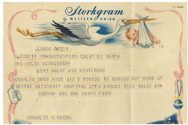
Iwas born on August 25, 1952 in Sacramento, California to Roberta Helen Rice and Charles Moen Rice Jr. (Fig. 1). My mother originated in Colorado Springs, Colorado, my father from Worcester, Massachusetts. I was the third Charles Moen Rice; we were all only children. I never met my paternal grandparents who were long gone by the time I was born. For most of my early years, we lived in Sacramento with a short interlude in San Jose (Fig. 2). My father worked as an insurance claims adjuster, my mother as a suburban housewife. My “brothers and sisters” growing up were a series of canine friends, largely dachshunds (Fig. 3). My parents and I loved dogs, a passion that endures to the present. A neighbor once said of my father, “if I believed in reincarnation, I would want to come back as one of your dad’s dogs.” No finer life could be imagined. I was an avid reader as a child, often staying up late into the night much to my parents’ chagrin. Music was a large part of my early home life nourished in part from my father who was a fanatic stereophile and had to have the latest high-end equipment. I was exposed to classical music and musicals, but later degenerated to listening to the Rolling Stones and the Doors on a low-end record player in my room.
Amongst my most cherished boyhood items were the usual chemistry set and very basic microscope, but my real love of biology and nature came from frequent camping and hiking trips in the Sierra Nevada mountains. My education was exclusively in public schools. I did reasonably well, despite having a series of odd jobs including cleaning up after parties at a nearby clubhouse. My custodial skills are still quite good. All in all, I had a fairly normal trajectory for someone growing up in the 1960s, which was anything but a “normal” decade: Vietnam, the cold war, the moon landing, assassinations of JFK, MLK and RFK, sex, drugs and rock-n-roll, etc.
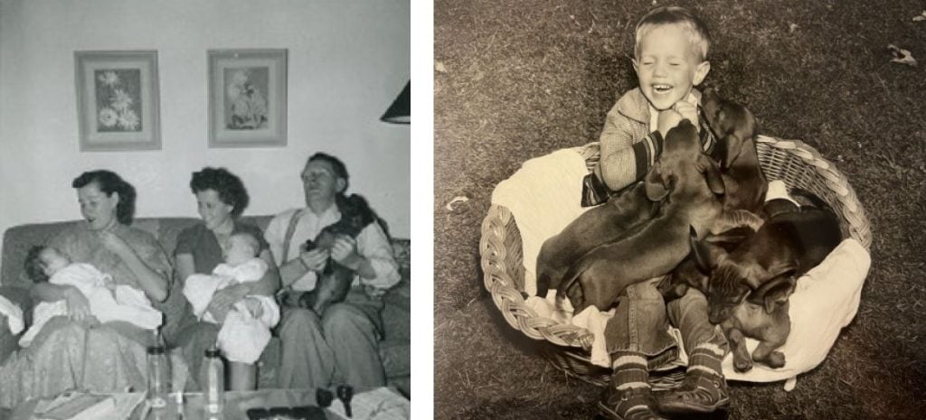
Figure 3. My “brothers and sisters”.
The last years of high school (Rio Americano), forced the choice of what to do next. The default trajectory for a middle-class kid was “go to college”, so that’s what I did. I applied to UC Davis and was accepted (with a small scholarship). For those of you unfamiliar with Northern California geography, Davis is about 15 miles west of Sacramento. So, for me, close to home, but far enough away. Given my love of animals, including even rats, I chose UC Davis given its veterinary medicine reputation, a vocation I thought I might pursue after finishing my undergraduate degree. In those days, going to a state university in your home state had an added bonus – it was cheap. I had no idea what major I was going to pursue but was leaning towards math until that was derailed by an advanced statistics course some years later. I blame this on my instructor, who had the appearance of wizard-like Gandalf, but the wisdom he was attempting to convey was lost on me.
An introductory biology course my freshman year turned out to be perhaps the most important chance encounter of my career. The course was taught by a Caltech-trained developmental biologist, Dennis Barrett. Dennis was/is a terrific biologist, and his lectures were captivating. Even more engaging were his exams, which were made up of multiple-choice questions. The exam was taken in class and the students could then take it home and revise their answers using any means available – textbooks, literature, or discussions with other students. This take-home portion of the exam, and the quest to get a perfect score, was an incredible learning experience because the correct answers to these multiple-choice questions were not the obvious ones and required a deeper dive and a real understanding of the biology. I don’t recall the details now, but Dennis became a key advisor and advocate for me during my undergraduate years and beyond.
My first undergraduate research experience was in the chemistry department studying tunicates (sea squirts) who manage to concentrate vanadium from sea water over six orders of magnitude in specialized blood cells. While I didn’t mind bleeding tunicates and doing elemental analysis, this wasn’t my cup of tea. I switched to working with Dennis on his favorite organism, the purple sea urchin, Strongylocentrotus purpuratas. “Purps” as we would call them are commonly found off the US Western coast; we collected them on field trips in the area near the Bodega Bay Marine Station. These animals can be induced to shed their gametes with an electrical jolt or injection of potassium chloride. Fertilization ensues by mixing the eggs and sperm in sea water, and you can watch early development proceed with a simple microscope – amazing. I was hooked, but this was just a part-time, sporadic research experience interspersed with working at the UCD library, the Memorial Union food service, and spending my first few summers working at a cannery or as a tomato inspector. It was some years before I could stomach eating ketchup again.
The summer of 1973 provided the chance to really immerse myself in research. Dennis was an instructor in the Physiology Course at the Woods Hole Marine Biology Laboratory (MBL) in Massachusetts and suggested that I apply to their summer program. This worked out. I arrived and found myself immersed in science 24/7. The course covered developmental biology, immunology, biophysical biochemistry and was taught by world class scientists through lectures, reading material and intense hands-on lab work. I turned 21 at the MBL and had a memorable (that I don’t actually remember) celebration (Fig. 4). After that, it was difficult to return to the undergraduate grind for my senior year at UCD.
Nonetheless, I survived and graduated in 1974 with a degree in Zoology. I’ve so far highlighted academic pursuits but in reality there was quite a bit going on outside of the classroom – soccer, softball, basketball, frequent backpacking trips and other activities that I will not detail here. Also in my academic wanderings, I had taken advantage of some “fun” classes, like the introduction to viticulture and enology anchored by the book “Wine: An Introduction” by UCD professors Maynard Amerine and Vernon Singleton. Growing up in California near the wine country, coupled with this course, seeded a real passion for wine. I found myself waffling between a career in wine and developmental biology research. Somewhat different trajectories …
Given this dilemma, I recruited a few college friends, we bought a used VW bus and departed for an extended trip to Central and South America. Before I left, I applied to two PhD programs strong in developmental biology: UC San Diego and Caltech. Uncertain as to my future, off we went, slowly making our way through Central America. After several months, a failing VW bus was abandoned in Costa Rica and we flew to Colombia to continue the South American leg of the adventure to Lima, Peru, where a telegram from my father awaited me: “you have been accepted at UCSD and Caltech and they want to know if you are coming”. I was having a great time traveling and meeting an amazing cast of wanderers, but a decision had to be made. Continuing from Lima to Cuzco, to Machu Picchu, La Paz and Salta in Northern Argentina, and after an exchange of letters with Dennis, I decided on Caltech, despite its lower stipend. A transition back to reality was needed. Fortunately, Dennis needed a teaching assistant for the MBL physiology course and some help moving from Davis to Denver, where he was joining the University of Denver faculty. The cheapest flight back to the US was to Miami. From there, an attempt by a scruffy vagabond with a backpack to hitchhike back to California failed, requiring a multiday Greyhound bus ride to Sacramento. After a few days back home, we loaded up a rental truck, drove from Davis to Denver and after a short stop to unload, headed to Woods Hole for another memorable summer. Besides the science, there was healthy competition between the various MBL courses, including a long-standing rivalry between the Physiology Course, headed by immunologist John Cebra, and the Embryology Course headed by the intensely competitive Eric Davidson, a Caltech professor who studied sea urchin development. This rivalry was played out on a softball diamond and a flag football field. My recollection is that the Physiology Course usually triumphed at softball whereas Eric’s team dominated the more physical football encounters. As you might have guessed, Eric’s sea urchin lab was one of the main reasons I was going to Caltech.
The summer ended and I was off to Pasadena, driving a yellow-orange 1975 VW Rabbit that my parents helped me buy. Being raised in Northern California, I approached this transition with great trepidation. How could I defect to Southern California? It was going to be smoggy, crowded, and terrible. My goal was to get my PhD and move on to greener pastures ASAP. But … I ended up loving it and spent nearly a decade at Caltech. However, there was a surprise upon my arrival. Rather than being assigned to Eric’s lab, as I’d hoped, I was placed in an animal virus lab, headed by James H. “Jim” Strauss and his wife Ellen Glowacki Strauss. In those days, laboratory rotations weren’t necessarily required or even encouraged. As I learned more about the Davidson lab I began to have second thoughts – Eric was brilliant but rumored to be tough, demanding and he didn’t suffer fools. I was pretty sure I belonged squarely in the “fools” category so I decided that this might not be the best fit. You could chalk it up to “no guts” and a lack of self-confidence, but at the same time the field of animal RNA viruses was still in its infancy and the Strauss lab was at the cutting edge of studying a model enveloped RNA virus, Sindbis virus, the prototype alphavirus. The lab was small with 3 PhD students, 2 techs, Jim and Ellen, and an excellent environment for someone entering a new field. Jim’s office was inhabited by two free-flying parakeets who didn’t necessarily take kindly to graduate students barging in for advice.
There had been extensive work in the lab on Sindbis virus RNA replication; less was known about the virus particle. Jim suggested that I work on a small, secreted glycoprotein called E3, which was processed from an envelope glycoprotein precursor during virion maturation and released into the cell culture supernatant. In the case of Sindbis, E3 was not virion associated whereas in the case of a related alphavirus, Semliki Forest virus, it stayed bound to the virus particle. I wasn’t enamored with this project. Fortunately, we were given a great deal of freedom to come up with and test our own ideas. I did end up doing quite a bit of work on the Sindbis virus glycoproteins, and that comprised the bulk of my thesis. But the larger Caltech research environment was playing a key role in shaping my research interests. This included a remarkable group of fellow PhD students and postdocs, the Caltech faculty and, of course, the undergrads. Recombinant DNA was exploding, we could now “easily” sequence nucleic acids and our lab was conveniently positioned right down the hall from the Maniatis lab, with its freezer full of enzymes and cutting-edge molecular biology expertise. Rather than being left out of this revolution, it seemed obvious that we should determine the sequences of the Sindbis structural proteins. The only catch was that concerns about recombinant DNA cloning and safety prevented molecular cloning of these animal RNA viruses. I was left to figure out how to sequence the structural protein coding region without complementary DNA (cDNA) cloning. Without delving into the gory details, it turns out that some restriction enzymes can cleave single-stranded cDNA or RNA-cDNA hybrids, providing discrete fragments that can be end-labeled, gel purified and sequenced using the laborious Maxim-Gilbert chemical sequencing method. This eventually provided the sequence of Sindbis subgenomic mRNA revealing a single open reading frame (ORF) that encoded the capsid protein followed by E3 and the two virion glycoproteins, E2 and E1. This capped off my PhD work, which I defended in 1981.
At this point, if not before, I should have been charting my future and lining up a postdoc. But to be honest, I was having too much fun collaborating with other labs, finishing ongoing work, and starting new projects. Again, this was possible because of the freedom granted, as long as good science ensued. I made a few half-hearted attempts, sending out feelers to Ron Davis at Stanford and Tom Cech in Colorado, but they either didn’t answer or weren’t interested (which is perhaps not surprising for an applicant working on an obscure RNA virus). So, ignoring the “academic kiss of death” I just stayed in the Strauss lab.
There were two ongoing efforts that made this an easy decision. The first revolved around an undying belief in the power of genetics. For me, this had been nurtured at UC Davis as an undergraduate by the great evolutionary geneticists Theodosius Dobzhansky and G. Ledyard Stebbins, and at Caltech surrounded by Max Delbruck in his waning years and the “next generation” including Bill Wood, Seymour Benzer, and Ed Lewis. One of the many attractions of working on viruses is their small genome size yet a highly evolved ability to infect cells, replicate and survive in nature despite a complete dependence on cellular functions. Inert on their own, they provide a perturbation to probe cell and organismal biology. As highly efficient machines, we can learn much from viruses themselves and their replicative mechanisms. Classically, chemical mutagenesis, selection of conditional mutants and their characterization under permissive and non-permissive conditions were the mainstay of animal RNA virus genetics. For Sindbis, this had certainly been the case for an immensely useful panel of temperature-sensitive (ts) mutants. But now, given reverse transcriptase and the ability to copy RNA into cDNA and recombinant DNA methods, how could we exploit these new tools to allow site-directed genetic manipulation of positive-sense RNA viruses?
In concept, this should be simple: isolate viral genome RNA, make a cDNA copy, and clone it into a plasmid that allows transcription of a functional viral genome RNA using either cellular machinery or in vitro transcription. This was shown to work for the bacteriophage QB by Taniguchi and Weissman in 1978. Shortly thereafter we began working towards the same objective for Sindbis. This was a collaborative effort with Henry Huang, a brilliant postdoc in Leroy Hood’s group. Cloning was now allowed and we were fortunate that Eugene Butler, a postdoc in the Maniatis lab, had purified and characterized the DNA-dependent RNA polymerase of Salmonella phage 6 (SP6) for his PhD and defined the 17-base promoter and initiating base. This turned out to be a highly efficient way to make unlimited quantities of synthetic Sindbis genome RNA with proper 5’ and 3’ terminal sequences. It took several more years, and additional work after I moved to St. Louis, before we published our first paper on the Sindbis infectious clone: “Production of infectious RNA transcripts from Sindbis virus cDNA clones: mapping of lethal mutations, rescue of a temperature-sensitive marker, and in vitro mutagenesis to generate defined mutants”. In the meantime, Vincent Racaniello and David Baltimore had reported success with poliovirus and Paul Ahlquist for the plant RNA virus, bromegrass mosaic virus. Many other examples followed, including recent work with SARS-CoV-2. This opened up an almost limitless landscape of possibilities for virologists since in theory, for any positive-sense RNA virus, these so-called “infectious clones” could be manipulated to generate mutations and phenotypes could be tested.
The second area cooking in the Strauss lab was an effort to understand more about another group of enveloped RNA viruses, the flaviviruses. Given their similar virion morphology and physical properties, alphaviruses and flaviviruses had been lumped together in the family Togaviridae. However, there was no serological cross-reactivity between these groups, so the flaviviruses had recently become a separate family, the Flaviviridae. Not much was known about flavivirus genome structure and how this might be similar or different from the alphaviruses or other positive strand RNA virus families.
The prototype flavivirus was the famous yellow fever virus (YFV), shown to be a filterable agent, transmitted by the bite of infected mosquitoes, and the cause of the often fatal viscerotropic disease by Walter Reed and his colleagues at the turn of the last century. Decades later, by passaging a human isolate from a young Ghanaian named Asibi in mouse and chicken embryo tissues, Max Theiler derived an attenuated yellow fever strain, called 17D, that was no longer virulent in non-human primates (NHP) and protected them from lethal YFV infection. This vaccine, which has been administered to more than 500 million people, is still used today. A single shot confers broad and long-lasting protection. Max, who was then working at the Rockefeller Foundation, was awarded the Nobel Prize in Physiology or Medicine in 1951 for his work on the yellow fever vaccine.
Given the choice of working with virulent flaviviruses like dengue virus, yellow fever virus or the Japanese, St. Louis, Murray Valley, West Nile and tick-borne encephalitis viruses, the YF 17D vaccine strain seemed like a no brainer. With great help from Edith Lenches, a stoic Hungarian technician in the Strauss lab, we spent years optimizing conditions for virus growth and partial purification of the virus. With high quality genome RNA in hand, and the assistance of three Caltech undergrads, we were able to clone and determine the sequence of YF 17D and, a few years later, the Asibi strain. Our YF sequences, together with the West Nile virus sequences reported by Gerd and Gisa Wengler, revealed a genome organization more like the picornaviruses than the alphaviruses – a genome of ~11 kb consisting of a single long ORF flanked by two short non-coding regions. Work from the Wengler lab had shown that flavivirus genome RNAs were capped but not polyadenylated. Rather, the new sequences revealed a potential stable 3’-terminal RNA secondary structure. This set the stage for a new era in flavivirus molecular biology, but what was needed were “infectious clones”.
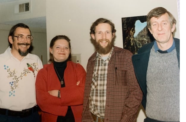
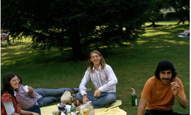
Given our ongoing Sindbis work, which was beginning to show promise, I decided to make the YF infectious clone effort a priority. Lynn Dalgarno from Australia was on sabbatical in the Strauss lab (Fig. 5) and we had been working together on another flavivirus, Murray Valley encephalitis virus. We became quite good friends and he extended an offer for me to spend some time in his lab at Australia National University. This seemed reasonable since it would allow me to really focus on the YF project. Before I left, I interviewed at a few places for a faculty position and received an offer from Washington University. Earlier, Henry Huang had joined their Microbiology and Immunology department (later changed to Molecular Microbiology). This was an especially attractive option since Henry and I had active Sindbis projects with Sondra Schlesinger in the Micro department. We had gotten to know Sondra well during her visit to Caltech to do some sequencing of Sindbis defective-interfering particle RNAs. I decided that Wash U was the best option for hitting the ground running, with the added bonus of not going out on any more job interviews. This was despite the not-so-generous start-up package of 60K offered by then department chair, immunologist Joe Davie, and a chorus of warnings from coast-biased colleagues about taking a position in the Midwest. This was surely “career suicide”.
Lynn warned me about the long lag time between ordering and receiving supplies at ANU so I packed up a complete collection of molecular biology enzymes and reagents before heading off to Canberra. My time in the Dalgarno lab was spent trying to assemble a full-length cDNA clone of YF 17D. I made literally hundreds of attempts but failed miserably. The reason: toxicity in E. coli. I did make some progress on learning which YF cDNA regions were tolerated versus toxic, which came in handy later. It was frustrating but the process was helped by frequent squash matches, an introduction to Aussie Rules football and a memorable stay on Heron Island, a beautiful coral gem on the Great Barrier Reef. There were some lasting upsides on the science front. With help from my future Wash U colleagues, we submitted a grant to the US Army to continue our YF efforts and I was selected to apply for the second class of the Pew Scholars Program. Fortunately, both were successful. Some years later, residual Pew funds helped jump start our work on hepatitis C.
Having survived left side drivers and a highly territorial Aussie magpie that swooped me whenever I wasn’t looking, it was time to return to Pasadena, pack up and head to St. Louis. The VW Rabbit, now 10 years older and showing its age, was loaded up with precious lab samples and my wine collection. I drove straight to St. Louis, acquiring two speeding tickets on the way. Besides setting up the lab, my first task was to write an NIH grant on the Sindbis work for the February 1986 deadline. It was painful (I wanted to be doing experiments), but I was excited: we finally had a functional Sindbis clone and I was able to borrow an Apple Macintosh 512K computer to help streamline the writing. What fun! Temperature sensitive mutants could finally be assigned to specific viral proteins, the Sindbis machinery could be engineered to express heterologous proteins rapidly and at high levels, and variants could be selected that were non-cytopathic in vertebrate cells.
The early years of the lab were a bit slow but productive. We “worked” all the time but also took time off to have fun outside the lab. I also met my life partner, Peggy MacDonald, and her chocolate Labrador retriever, Bonnie. Most of the first crop of PhD students worked on Sindbis projects, I continued slogging away on YF 17D. I tried installing dual transcription terminators at the 5’ and 3’ YF-plasmid boundaries, low copy plasmids, different E. coli hosts, and finally I just abandoned plasmids altogether and tried lambda phage. I figured that if lambda was going to lyse the bacterial host anyway, maybe flavivirus cDNA toxicity wouldn’t be an issue. Not so – it did work but the recombinant phage was unstable and the burst size was so small that DNA yields were too low to be useful. The polymerase chain reaction was seeing increased use but in those days the error rate was high, and it was difficult to amplify long templates. From my failed attempts in Australia and at Wash U, I knew that the full-length cDNA could be propagated in E. coli as two plasmids that could be designed to overlap by unique restriction enzyme sites. Why not try assembling the full-length template for SP6 transcription by in vitro ligation and completely avoid trying to amplify the full-length YF cDNA in bacteria? This took some optimization, but it worked. A modern version of this strategy is still used today to assemble flavivirus and coronavirus DNA templates for in vitro transcription of full-length genome RNAs.
Energized and hopeful, we sent the paper off to Science. As I recall, it went through 2 rounds of reviews that were largely favorable, but it was ultimately rejected because of a perceived “lack of general interest”. A new journal, The New Biologist, offered to publish the work and it finally appeared in 1989. The New Biologist perished a few years later, making these reprints a real collector’s item. I don’t have any, so don’t bother contacting me.
Nonetheless, we were finally poised to explore the molecular genetics of YFV. The top priority was to understand the molecular basis of the attenuated YF 17D vaccine strain. This would involve making an Asibi infectious clone, testing the resulting virus for virulence in rhesus macaques, and then making a series of 17D/Asibi recombinant viruses to map the changes responsible for attenuation. This was a collaboration with Joel Dalrymple at USAMRIID who had made the Asibi RNA that we used for cloning and sequencing. Joel would undertake the NHP work, my lab would make and characterize the chimeras in cell culture models and make stocks for animal inoculation. Tragically, Joel developed aggressive kidney cancer and passed away a few years later at the age of 53. Without our USAMRIID collaborator and internal advocate, the project stalled and remains unfinished to this day.
Other work on YF was progressing. We determined the order and boundaries of the mature flavivirus proteins and began to unravel the complex proteolytic processing scheme involved in their production. One key player was a viral serine protease embedded in the polyprotein that mediated cleavages required for assembly of infectious virus and the membrane associated RNA replication machinery. We could show that this protease activity was essential, as were each of its seven site-specific cleavages in the YF polyprotein.
At the time, we were not working on non-A, non-B post-transfusion hepatitis. From work in the chimpanzee model, the mystery agent appeared to be a small, enveloped virus, but its true identity was unknown; attempts to culture it in the laboratory had failed. Unexplained cases of hepatitis were on my mind, however, given the death of a close friend’s mother from acute liver failure, cause unknown.
This changed shortly after the 1989 publication of landmark papers from Michael Houghton, Harvey Alter and their colleagues, identifying a new positive-strand RNA virus (called hepatitis C virus, HCV), with a genome of about 10 kb, as the cause of non-A, non-B post transfusion hepatitis. HCV infection was widespread, usually chronic and associated with mild to advanced liver disease, including cirrhosis and cancer. The sequence of the virus revealed a single long open reading frame encoding a polyprotein with features strikingly similar to members of the Flaviviridae, including the classical flaviviruses and the animal pestiviruses. A new virus member had joined our family.
As a relatively new Assistant Professor, it didn’t make much sense to start working on a virus that couldn’t be grown in the laboratory. This brief period of sanity was derailed by a phone call from Steve Feinstone at the FDA who had read our YF 17D infectious clone paper. Steve wondered if this couldn’t be leveraged to produce a vaccine against HCV. Steve, a co-discoverer of HAV, had been characterizing the non-A, non-B agent in the chimpanzee model and, like many others, was attempting to identify the causal virus.
With Steve’s nudge and encouragement, we decided to collaborate and started working on HCV, with the initial goal of learning more about HCV polyprotein processing and the functions of the resulting mature viral proteins – very similar to what we had done for YF. Steve had access to some of Harvey Alter’s famous patient H plasma obtained in 1977 during the acute phase of infection and shown to be highly infectious in chimpanzees. The first step was to make HCV H-strain cDNA clones for expression studies in mammalian cells and in bacteria, to make regionspecific antigens, and a panel of HCV-specific antisera. The problem? No one in the lab wanted to work on HCV. Generating antisera was boring and besides, there was no “virus” to work with in the lab.
Arash Grakoui (Fig. 6) stepped up to the plate in my lab, Steve and his team pitched in and together we managed to generate a near complete map of the HCV polyprotein cleavage products, identify two HCV-encoded proteases and their cleavage sites, and start some collaborative work on other predicted HCV enzymes. We weren’t alone. Other groups in the US, Europe and Japan were reporting similar results and the HCV research community was growing. There was a sense of the field’s importance, a mutual respect for our colleagues and competitors, and open sharing of ideas and materials. This nurturing atmosphere was seeded in large part by Michael Houghton, who initiated and anchored a yearly international HCV meeting where he would highlight and applaud every advance in the field.

But the central problem persisted. Advances were being made, but we still didn’t have an HCV permissive cell culture system. I seemed to be on a downward spiral, starting with a virologist-friendly alphavirus, then a more challenging flavivirus and now, a virus that we couldn’t even grow in the lab. We began to work on the animal pestiviruses, the closest relatives to HCV at the time, and which replicated in cell culture. This provided some needed experimental “relief”, but we hadn’t given up on HCV.
Early on, we had decided to try a “reverse” strategy. If we couldn’t get infectious patient or chimpanzee serum to infect cells in culture, perhaps we could raise our chances by using infectious clone-derived HCV RNA. We would have an unlimited supply of infectious material to screen for permissive cells and conditions. The dilemma was how to assay infectivity – there was only the chimpanzee model and intra hepatic injection of a 9.6 kb RNA and hoping it would be taken up by hepatocytes, intact, seemed like a stretch. It wasn’t completely far-fetched though, given that Susan Emerson and Robert Purcell had successfully taken this approach years earlier for hepatitis A virus RNA in marmosets.
We assembled what we believed was a full-length cDNA clone from Harvey Alter’s H77 material, made RNA and Steve injected this intrahepatically into chimpanzees. Nothing happened – no evidence of HCV RNA in circulation but we did learn the input RNA was very rapidly degraded and quickly became undetectable. Disappointing but perhaps not too surprising given this crude and likely inefficient RNA transfection attempt.
At this time, we began to wonder about our full-length clone. Was it really full-length, or might there be something missing? HCV sequences were being reported around the world and while the original sequence reported by the Houghton group terminated with polyA, the subsequent sequences suggested polyU. A careful analysis of the cDNA cloning methods used suggested that they could be biased. Alexander “Sasha” Kolykhalov, a superbly talented molecular biologist who had immigrated from Russia, took on the challenging task of confirming or extending the 5’ and 3’ terminal sequences of the HCV genome. Had Sasha not joined my group, I doubt that I would be writing this piece. Immigrants have and continue to make some of the most impactful contributions to US and global success stories. Isolating ourselves from this flow of untapped talent is a sure formula for disaster.
I will keep the next specifics to a minimum since they are covered in my Nobel Lecture. Sasha went on to discover a missing piece at the 3’ end of the HCV genome RNA. This later turned out to be absolutely required for virus replication. However, modifying our existing clones with this new sequence still failed to initiate productive replication in chimpanzees. Why was this? We worried about everything. Missing sequences, transfection method, a requirement for RNA modifications or a viral protein for infectivity, lethal mistakes in our clone, and more. The list was daunting. Sasha cloned and assembled hundreds of full-length cDNA clones, tested them with restriction enzyme digestions and in culture for polyprotein translation. He sequenced a subset that passed these criteria and used this information to assemble a clone that reflected the dominant H77 consensus sequence. When Steve inoculated this RNA into two animals, we saw a rise in circulating HCV RNA, characteristic liver inflammation and delayed seroconversion; both animals went on to chronic infection. After eight years of working on HCV, we finally had an infectious clone; more examples followed soon thereafter. The journal Science did accept this one.
Despite being validated in vivo, transcripts from HCV consensus clones were unable to replicate in cell culture. Another breakthrough, another roadblock. Many laboratories tried different HCV isolates, diverse cell types and conditions, but nothing worked. The ability to launch infection in the chimp model opened up new possibilities to study HCV evolution, immunity and test the impact of mutations on virus viability but the cell culture roadblock remained. Overcoming this was important, not just for studying the virus lifecycle but also for developing antiviral drugs. Without a cell-based assay supporting virus replication, potent inhibitors could be developed using biochemical assays but there was no convenient pre-clinical assay to test and optimize their efficacy at blocking virus replication.
The next breakthrough, reported in Science in 1999, was from Ralf Bartenschlager’s lab at the University of Mainz in Germany. Like many in the field, Ralf and his student, Volker Lohmann, were close to quitting their efforts on HCV and moving on but decided to try one more last-ditch effort. Starting with a consensus clone for a German isolate, they engineered an HCV RNA where the portion of the ORF encoding the virus structural proteins was replaced by the gene for neomycin resistance. This was followed by an internal ribosome entry site from encephalomyocarditis virus (EMCV) to drive translation of the HCV proteins needed for viral RNA amplification. If this engineered RNA could replicate and express the drug resistance gene, then cells would be resistant to the drug and able grow in its presence. It worked – a few drug-resistant colonies appeared and the HCV subgenomic replicon system was born. We had been trying a similar approach with our H77 infectious clone but failed to detect any drug-resistant colonies. In need of a positive control, we assembled the Bartenschlager subgenomic replicon using synthetic oligonucleotides and confirmed their results. Remarkably, when we and others sequenced the HCV RNA present in these rare colonies, it was not the sequence of the input RNA. When even single changes were engineered back into the original replicon sequence, some of them raised the efficiency of drug resistant colony formation by more that 10,000-fold. Ten years after HCV’s discovery, we finally had a robust cell culture system for studying RNA replication and aiding drug development.
At this point, another phone call ushered in a major change in my life. Steve Goff, a virologist at Columbia, was on the line asking if I would visit Rockefeller University and advise a committee tasked with finding someone to head a new Tri-Institutional “Center for the Study of Hepatitis C”. One thing led to another, and I was offered the position. But I was perfectly happy at Wash U. My stint as interim chair of Micro was finished so I could really focus on research, and, being an outdoor, non-city type, I had always done my best to avoid New York City. Not to mention that Rockefeller, with its stratospheric reputation, didn’t seem like the right landing pad for a plodding molecular virologist. After some tough soul searching and a nudge from my scientific grandfather, Jim Darnell (you should do this!), Peggy and I decided to make the move. Peggy gave up her tenure track faculty position at Wash U, and we moved to NYC in mid-2000 to an apartment that was about 50 yards from the Rockefeller campus. Our two rescue dogs, Sadie and Wrangler, were not happy with the loss of their yard and the new digs. But we all managed to adjust aided by the vibrant scientific atmosphere at Rockefeller, wonderful colleagues who, after all, were not that scary, and a beautiful park-like campus.
Not many of the Wash U crew were able to relocate from St. Louis to NYC so the first years were spent rebuilding the group, closing the Wash U lab and waiting for our renovated space in the RU Hospital building to be finished. I was fortunate to have a few previous trainees return to the lab for postdocs as well as two senior scientists who joined the Center and formed groups working on HCV immunology and virus entry (using pseudovirus approaches since we still lacked cell culture infectious HCV). We also solidified interactions with clinical colleagues and the Clinical Director of the Center, Ira Jacobson, who is a renowned clinical hepatologist working at the time at Weill-Cornell and New York Presbyterian Hospital. Ira shared over the years a wealth of knowledge regarding important aspects of HCV diagnosis, treatment, and challenges that HCV infected individuals face, helping to keep the ultimate goal of the research clearly in sight.
From the HCV replicon work, the next step was obvious but when adaptive mutations were placed in full-length, otherwise unmodified HCV RNAs, they replicated and expressed the expected HCV proteins, but no virus was produced. A major advance, another roadblock. This became yet another annoyance for the field that persisted for another 5 years. This was finally solved by a serendipitous finding in Japan by Takaji Wakita. Takaji was studying an HCV isolate from a rare case of acute fulminant disease (Japanese fulminant hepatitis 1, or JFH1). Perhaps this virus, given its atypical pathogenesis in the patient might be able to replicate in culture. When he tested a JFH replicon, it replicated efficiently in human hepatoma cells with no need for adaptation. Later work by three groups, including ours, showed that JHF1 or chimeric HCV derivatives could produce virus that was infectious in cell culture, chimpanzees, and mice engrafted with human hepatocytes. More than 15 years after HCV’s discovery, we finally had a complete cell culture replication system.
It was another six years before the first HCV protease inhibitors entered the clinic and several more years before IFN was abandoned in favor of effective drug cocktails that could eliminate the virus in virtually everyone treated, with minimal side effects. In the intervening years, much was learned about the intricacies of HCV RNA replication, virus entry, and the host cell factors important for virus infection. Efforts ramped up in the biotech and pharma sectors working to identify and optimize drugs for inhibition, to identify resistance mutations and their effect on viral fitness, and to test combinations of drugs for efficacy against the diversity of HCV genotypes present in the infected population and to determine the minimal duration to achieve a cure – elimination of the virus from an infected individual! The ultimate outcome, combination drug cocktails capable of achieving virological cure in two to three months across multiple HCV genotypes, was an amazing culmination of the hard work of so many individuals, across a diverse spectrum of fields. Looking back, I feel incredibly fortunate to have had, by a series of chance events in my life, the opportunity to be a part of this effort, and to see first-hand the role that basic science can play towards helping patients afflicted with a devastating disease. Not every basic scientist will be so lucky to be in such a “right place at the right time”, but the future cannot be predicted, and random chance could strike at any time. I urge all young (and not so young) scientists to embrace their passion, follow their curiosity where it leads them, work hard, and to be fair and open in sharing ideas and reagents. No one discovery is likely to change the world, but the collective work of all of you will.
© The Nobel Foundation 2024
Nobel Prizes and laureates
Six prizes were awarded for achievements that have conferred the greatest benefit to humankind. The 12 laureates' work and discoveries range from proteins' structures and machine learning to fighting for a world free of nuclear weapons.
See them all presented here.

